

 PDF
PDF ePub
ePub Citation
 Print
Print


Primary angle-closure glaucoma (PACG) is an anatomical disease in which the peripheral iris contacts the trabecular meshwork and thus blocks the access of aqueous into it. Due to the increased intraocular pressure (IOP) caused by the blockage of the aqueous outflow, glaucomatous optic nerve damage occurs. If the pupillary block is marked, pronounced iris bombe leads to complete appositional obstruction of the trabecular meshwork and thus IOP is increased rapidly resulting in the development of acute angle-closure glaucoma (AACG) that is accompanied by ocular pain, headache, and decreased visual acuity. On the other hand, chronic angle-closure glaucoma (CACG) is the glaucoma in which the peripheral iris is in apposition to the trabecular meshwork with a gradual progression of the adhesion and consequently IOP is raised gradually, with subjective symptoms such as ocular pain, haloes, etc., therefore being only rarely present.
The major mechanism of PACG is believed to be the pupillary block, however, in many cases, the pupillary block is not the only mechanism. For example, the plateau iris induces the angle crowding regardless of the pupillary block, and the angle crowding may also exist owing to an anteriorly positioned thick lens. Another mechanism of angle closure is ciliochoroidal effusion, and expansion of the choroid may aggravate both pupillary block and angle crowding.1,2
Peripheral iridotomy for AACG and CACG is effective for to widen the filtration angle and reduce the increased IOP.3 In some instances, persistent IOP elevation may occur despite a patent iridotomy because of trabecular damage from repeated subacute angle-closure attacks or from significant synechial angle closure. The pigment granules and the iris tissue fragment released during laser peripheral iridotomy may exacerbate the aqueous outflow of the trabecular meshwork that has been damaged already. In addition, even if iridotomy were performed, the lens located relatively anteriorly or the thick lens may cause the appositional angle closure.4,5
Greater extent of peripheral anterior synechiae, a higher IOP before surgery, and a larger cup to disc ratio have been reported to be poor prognostic factors in controlling IOP.6,7 If severe synechial angle closure and glaucomatous optic nerve damage are present, the effect of IOP reduction after laser peripheral iridotomy is small, and thus it is difficult to protect the optic nerve. Hence, in PACG, early surgery has been recommended in the presence of severe glaucomatous optic nerve damage and damaged visual field at presentation, or the presence of 50% to 75% of the angle closure by peripheral anterior synechiae on indentation gonioscopy.7-9 Therefore, the status of the optic nerve head has an important implication to assess the prognosis of PACG.
It has been reported that the correlation between pretreatment IOP and the amount of visual field damage is higher in PACG than in primary open-angle glaucoma, and that the cup to disc ratio is correlated with the severity of visual field damage. Hence, in PACG, glaucomatous optic nerve damage is associated primarily with high IOP. However, in primary open-angle glaucoma, other factors in addition to increased IOP may be related.10 In PACG, high IOP is an important risk factor for the development of glaucomatous optic nerve damage. Therefore, in AACG, with high IOP caused by the acute attack, and in CACG, with the moderate level of increased IOP for a long time, we investigated any differences in glaucomatous optic nerve damage between the two groups.
Materials and Methods
The study included 84 normal individuals, 130 AACG patients with acute attack, and 86 CACG patients without acute attack. The PACG patients who visited the department of ophthalmology at Hanyang University Hospital from 1993 to 2003 were examined by assessing the qualitative signs of glaucomatous optic nerve damage to determine whether optic nerve damage differs according to the presence or absence of acute attack. Criteria for the diagnosis of AACG in this study were as follows: an acute attack accompanied by high IOP elevated over 40 mm Hg that may induce iris tissue ischemia or corneal edema, ocular pain, red eye, decreased visual acuity, nausea, vomiting. A closed angle on gonioscopy and the cases with or without glaucomatous visual field damage were included. Criteria for the diagnosis of CACG in this study were as follows: no history of acute attack, an elevated IOP over 21 mm Hg, angle-closure with peripheral anterior synechiae, and cases with or without glaucomatous visual field damage. To avoid any selection bias, the appearance of the optic disc was not taken into account for diagnosis. The study subjects were classified according to the classification proposed by Kim and Jung,11 as shown in Table 1. The cases of secondary angle-closure glaucoma caused by phacomorphic glaucoma, neovascular glaucoma, and iridocyclitis were excluded from the study subjects. Normal eyes had no lesions in the anterior or posterior segment other than dry eye, incipient cataract, and refractive error. The maximum corrected visual acuity was over 0.7, family history of glaucoma was absent, IOP measured by Goldmann applanation tonometer was lower than 21 mm Hg, and normal findings were shown in central 30-2 full threshold strategy by Humphrey visual field analyzer (Allergan Humphrey, San Leandro, CA., U.S.A.). Among the normal eyes of 181 individuals who were examined for qualitative signs of glaucomatous optic nerve damage, age, gender, and refraction in the normal eyes of 84 were matched to those of the AACG and CACG groups and these 84 eyes were designated as the control group.
The severe myopia cases with spherical equivalence more than -8 diopters and the astigmatism cases with more than 3 diopters were excluded from the study subjects. In the cases of normal eyes and both eyes having glaucoma, one eye per individual was selected randomly for statistical analysis.
After the dilatation of the pupil, 20-degree color polaroid photographs of the optic disc were taken using a fundus camera (Topcon TRC-50X or TRC-50EX, Japan). Blurry pictures were excluded from the study population. The presence or absence of the following ten qualitative signs for differentiating between normal and glaucomatous optic discs were recorded: seven optic disc findings of an abnormally shaped neuroretinal rim width (alteration of ISN'T rule), neuroretinal rim width narrower than the temporal sector, rim notch, disc hemorrhage, bared circumlinear vessel, vessel bayonetting, and vessel nasalization, and three peripapillary findings of abnormally large peripapillary atrophy, abnormal form of peripapillary atrophy, and zone beta. Optic disc was divided into 60-degree temporal horizontal sector, 90-degree superior temporal sector, 90-degree inferior temporal sector, and 120-degree nasal sector. In all sectors, the presence or absence of the neuroretinal rim width narrower than the temporal sector, the abnormal form of peripapillary atrophy, and the zone beta were recorded. The border of the optic nerve head was the inner side of the scleral ring, the cup margin was the contour line referring to the shape of the blood vessel bend, and the vertical cup to disc ratio was measured using a ruler. If the neuroretinal rim tissue was absent under the blood vessel, it was considered as the optic cup. The zone beta surrounding the optic disc was evaluated as a time unit with up to 0.5 hour unit being measured.
'Abnormally shaped neuroretinal rim width' was defined as the cases of the inferior sector of the neuroretinal width being narrower than the superior sector, the superior sector being narrower than the nasal or temporal sector, and the nasal sector being narrower than the temporal sector. 'Neuroretinal rim width narrower than the temporal sector' was defined as the thinnest neuroretinal rim width in other regions being narrower than the minimal neuroretinal rim width within 60 degrees of the temporal horizontal sector.12,13 'Rim notch' was defined as a 60-degree sector of the optic disc in the center of which the neuroretinal rim was thinner than 50% of the rim width at both peripheral borders of the disc sector. 'Disc hemorrhage' was defined as a splinter shaped or dot like hemorrhage in the neuroretinal rim or in the margin of the optic disc. Circumlinear vessels are small branches arising just above or below the bifurcation of the central retinal artery or vein that follow a curved path along the margin of the optic cup and heading toward the macula, however, the cases with this blood vessel located not in the cup margin but in the cup area were designated as the 'Bared circumlinear vessel'.13,14 'Vessel bayonetting' was defined as the cases of the retinal vessel made a sharp bend as it crosses the cup margin. 'Vessel nasalization' was arbitrarily defined as the cases of the major retinal vessel being displaced nasally more than half of the horizontally connected line from the center of the optic disc to the nasal optic disc margin. 'Abnormally large peripapillary atrophy' was defined as the cases of the zone alpha or zone alpha and beta having a maximum width of more than 1/5 of the horizontal optic disc diameter. 'Abnormal form of peripapillary atrophy' was defined as the cases of the maximal extension, regardless of the atrophy size, not being arranged symmetrically around the horizontal but rather directed towards the inferior temporal or superior temporal region, or located in the nasal sector.13 'Zone beta' was designated as the peripapillary atrophy adjacent to the optic disc in the area showing choroidal blood vessels or the sclera.15
Glaucomatous visual field damage on the Humphrey visual field analyzer was defined as a reproducible loss of 10 dB or greater in two or more contiguous points in Bjerrum's area, a reproducible loss of 5 dB or greater in three or more contiguous points in Bjerrum's area, or the appearance of a 10 dB difference across the nasal horizontal midline in two or more adjacent locations.9 The study subjects comprised the cases with fixation loss of less than 20%, and with false positive and false negative response of less than 33%. The cases with eye diseases possibly affecting the vision, a history of eye trauma, optic nerve lesions other than glaucoma, and opacity of the ocular media were excluded from the study subjects.
The continuous variables of the normal, AACG, and CACG groups were compared by one-way ANOVA. For categorical variables, Chi-square test or Fisher's exact test was used. To assess the qualitative signs of optic nerve damage that are diagnostically valuable in AACG and CACG groups, specificity and sensitivity were evaluated. P values less than 0.05 were considered significant.
Results
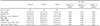
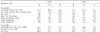
The mean age of the normal, AACG , and CACG groups was 62.1, 61.3, and 64.0 years, respectively, and no difference was detected between the normal and AACG groups, or between the normal and CACG groups. However, the CACG group was significantly older than the AACG group (p=0.02). The proportion of females was 69.0%, 81.5% and 75.6%, respectively, and the differences were not significant. The average refractive error was 0.46, 0.58, and 0.30 diopters, respectively, and the differences were not significant. The average mean deviation (MD) was -10.2 dB in AACG group and -13.1 dB in CACG group, and the difference was significant (p=0.04). The average corrected pattern standard deviation was 4.1 dB in AACG group and 5.1 dB in CACG group (p=0.05). The average cup to disc ratio in the normal, AACG and CACG groups was 0.47, 0.64, and 0.74, respectively, and all were significantly different (p<0.05). The average zone beta surrounding the optic disc was 0.59, 2.34, and 3.56 hours, respectively, and all were significantly different (p<0.05) (Table 2).
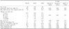
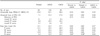
In the normal, AACG, and CACG groups, the frequency of the abnormally shaped neuroretinal rim width (alteration of ISN'T rule) was 28.6%, 60.8%, and 81.4%, respectively (p=0.001). Bared circumlinear vessel was detected in 10.7%, 34.6%, and 54.7% (p=0.004), vessel bayonetting in 4.8%, 15.4%, and 30.2% (p=0.009), neuroretinal rim width narrower than the temporal sector in 4.8%, 43.8%, and 58.1% (p=0.04), zone beta in the nasal sector in 1.2%, 11.5%, and 22.1% (p=0.04), and zone beta of the superior temporal sector in 8.3%, 29.2%, and 43.0% (p=0.04), respectively. These findings were detected significantly more frequent in CACG group than in AACG group (Table 3, 4). In AACG and CACG groups, the most useful qualitative sign was the abnormally shaped neuroretinal rim width. The specificity and sensitivity of the abnormally shaped neuroretinal rim width was 71.4% and 60.8% in AACG group, and 71.4% and 81.4% in CACG group, respectively (Table 5).
Discussion
It has been reported that in an average 6-year follow-up of Asian AACG patients, glaucomatous optic nerve damage was detected in almost half, and the cup to disc ratio was over 0.9 in 16%. In addition, due to the ischemic change of optic nerve, an optic disc pallor larger than the glaucomatous cupping was detected in 20%.16 In AACG, including the cases without optic nerve or visual field damage, an average 4-year follow-up after laser peripheral iridotomy showed that IOP was increased after the recovery of the attack in 58.1%, and thus medications were required, and that IOP was elevated within 6 months in 76.6%. Filtering surgery was required eventually in 32.7% because of poorly controlled IOP.17 Such results suggest that for AACG, laser peripheral iridotomy alone is not sufficient as a long-term treatment. Reported possible reasons are delayed visits to the eye clinic, very high IOP, a plateau iris, angle crowding by the lens, pigment dispersion and inflammation caused by laser peripheral iridotomy, and the difference of the clinical outcome between Asians and Caucasians.17-19
The prognosis of the visual loss in AACG is associated with the duration and severity of acute attack. A long duration of acute attack and very high IOP may cause permanent damage to the optic nerve.20,21 However, in AACG, the visual field damage may be developed due to various factors, in addition to acute attack, such as previously existing CACG, previously frequent subacute attack that was recovered spontaneously, and chronic increased IOP after the recovery of acute attack. If CACG is developed after acute attack, IOP is high, control with medication is difficult, the progression is very fast, and, hence, surgery is required frequently.17,22
CACG is a disease prevalent in Asians. Characterized by synechial closure of the filtration angle as a result of iris apposition to the trabecular meshwork, it progresses gradually asymptomatically, and filtration angle is closed permanently. Iridotomy is performed to treat the papillary block. Nevertheless, iridotomy alone is not sufficient in many cases and eye drops are required frequently to lower IOP. After iridotomy, the mechanisms, other than pupillary block, causing the elevation of IOP are the plateau iris, lens factor, damaged trabecular meshwork function, etc. Because of the elevated IOP, glaucomatous optic nerve damage appears eventually.23,24 In CACG, high pretreatment IOP has been shown to be associated with the peripheral anterior synechiae. In addition, pretreatment IOP is associated with the cup to disc ratio, which has been reported to result in IOP being an important risk factor in CACG for the development of glaucomatous optic nerve damage.25 It has been reported that for peripheral anterior synechiae over 180 degrees, IOP is increased in most cases, and that if the filtration angle is closed in over 270 degrees, medications are ineffective and filtering operation is therefore required.26
Dhillon et al27 have reported that the visual field damage is more frequent in CACG than in AACG, and Ang et al28 have reported that the visual field damage is more severe in CACG than in AACG. In addition, the cup to disc ratio is higher and the damage of the nerve fiber layer is more severe in CACG than in AACG.29,30 Choi and Kim31 have reported that the correlation of the peripheral anterior synechiae and the visual field damage was higher in angle-closure glaucoma without attack than in angle-closure glaucoma with attack.
In our study, different from previous analyses, the frequency of the qualitative signs of the glaucomatous optic nerve damage was examined. The results show that abnormally shaped neuroretinal rim width (alteration of ISN'T rule), bared circumlinear vessel, vessel bayonetting, neuroretinal rim width narrower than the temporal sector, and zone beta in the nasal and superior temporal sectors were more frequent in CACG group than in AACG group. In addition, the MD of the visual field, vertical cup to disc ratio, and extent of zone beta were significantly more severe in CACG group than in AACG group. Therefore, our results support the results reported previously that glaucomatous optic nerve damage appears to be more severe in CACG than in AACG, and it was thought that the optic nerve is better able to endure short-term, high IOP than long-term, moderately elevated IOP. In CACG, the characteristics of the disease are an insidious nature with the absence of any dramatic visual symptoms. As this makes it difficult for patients themselves to find the abnormalities despite evident glaucomatous optic nerve damage, it is thought that the diagnosis and treatment may be delayed in many cases. On the other hand, it is thought that in AACG, due to the acute nature of the attack, patients and clinicians may be aware of the presence of the disease, allowing treatment to be administered relatively rapidly and thus severe glaucomatous optic nerve damage to be prevented. In addition, it has been reported that in AACG, although the severe damage of visual function that is caused by the acute attack may be induced for a short-time, in many cases it is recovered without visual loss and, therefore, glaucomatous optic nerve damage is more severe in CACG than in AACG.21,32,33 Ang et al28 have reported that the mean age of AACG and CACG was not different, although in our study the mean age of the CACG group was higher. Additional studies may be required to determine whether the age of the CACG group was higher because of the longer duration of the elevated IOP and the later diagnosis. In our study, in both AACG and CACG groups, the most useful qualitative sign was the abnormally shaped neuroretinal rim width, which has also been reported to be the most useful finding in primary open-angle glaucoma.34
Greater extent of peripheral anterior synechiae, a higher presenting IOP, a larger cup to disc ratio have been reported to be poor prognostic factors for the control of IOP after iridotomy.6,7 Salmon6 has reported that in CACG cases that had undergone laser peripheral iridotomy, the best prognostic marker to decide whether or not to perform filtering operation is the cup to disc ratio. In AACG, even if IOP is normal during the initial period after iridotomy, asymptomatic, chronic elevation of IOP could occur within one year,35 and even if medications were effective on CACG, the progression is rapid in comparison with primary open-angle glaucoma.10 Therefore, it is very important in AACG and CACG to examine thoroughly whether IOP is increased, to determine whether the peripheral anterior synechiae and optic nerve damage is progressed, and to administer appropriate treatments.
In conclusion, it appears that glaucomatous optic nerve damage is larger in CACG without acute attack than in AACG with acute attack. It is thought that in PACG, it is the duration, rather than the degree, of the IOP elevation that appears to mediate primarily the effect on glaucomatous optic nerve damage, which suggests that the optic nerve is better able to endure short-term, high IOP than long-term, moderately elevated IOP.
 XML Download