

 PDF
PDF ePub
ePub Citation
 Print
Print


Loss of volume after enucleation of the globe is the primary cause of cosmetic problems of the orbit. Among various problems, the correction of superior sulcus deformity and enophthalmos in the anophthalmic orbit is one of the most challenging part to oculoplastic surgeons. Even a large prosthesis can't solve the undesirable appearance of depression of superior tarsal area and enophthalmos. Most often, in addition to these major cosmetic problems, ptosis of the upper eyelid and stretching of the lower eyelid are also present. These total appearances are called "anophthalmic orbit syndrome" or "postenucleation socket syndrome".1,2 There also can be shown upper eyelid retraction(in contrast to upper eyelid ptosis) with deepening of the sulcus because of retraction of the superior muscle complex(superior rectus and levator muscle).3
Several etiologies of anophthalmic orbit syndrome have been postulated: 1) atrophy of orbital fat,4 2) migration of muscle cone,5 3) traumatic bony loss, 4) herniated orbital fat secondary to unrecognized orbital wall fracture,6,7 5) loss of volume when globe is removed,8 6) levator disinsertion,9 7) malposition of superior rectus muscle, 8) long-standing gravitational burden of orbital implant and prosthesis.
Superior sulcus deformity and enophthalmos may happen even after placement of an adequately sized orbital implant. Furthermore, prosthesis placement alone is often not adequate for the volume loss in enucleated socket. A variety of autogenous and alloplastic materials such as glass beads,10 autologous or homologous bone,4 cartilage,11 dermis-fat grafts,12 plastic plate,13 room-temperature vulcanized(RTV) silicone,14 synthetic porous composite of Teflon polymer and alumina (Proplast II)15 have been used as subcutaneous or subperiosteal implant to correct superior sulcus deformity and enophthalmos.
Considering long standing gravitational forces of orbital implant and prosthesis, subperiosteal implant has some several advantages over placing autogenous or non-autogenous materials directly in the eyelid to fill the defect of superior sulcus. Porous high-density polyethylene (Medpor®) implants are useful because they are easily inserted into the subperiosteal space and provide appropriate long standing volume augmentation to push the orbital contents superiorly and anteriorly.
To estimate the effectiveness of correcting superior sulcus deformity and enophthalmos, Medpor® sheets were used as a subperiosteal implant in a total of 11 anophthalmic patients in this study. The indication for this procedure was patients who showed superior sulcus deformity and enophthalmos despite the prior insertion of an adequately sized orbital implant and prosthesis.
Materials and Methods
The study was designed retrospectively to estimate long term effectiveness of subperiosteal Medpor® sheet implantation in anophthalmic orbit. We reviewed hospital records of 11 anophthalmic patients who underwent volume augmentation surgery with Medpor® sheet between August 1997 and March 2004. The patients were selected for surgery on the basis of poor cosmesis owing to the presence of a deep upper eyelid sulcus and enophthalmos despite the presence of an adequately sized orbital implant and prosthesis.
Photographs were taken in all patients to compare degree of superior sulcus deformity and exophthalmometric value with their own prosthesis was assessed to measure enophthalmos using Hertel exophthalmometer preoperatively. The volume and structure of the implant, surrounding extraocular muscles, soft tissue and possible orbital wall fracture were carefully examined by using computerized tomographic scans and ultrasonography in some cases.
In 8 of 11 patients, volume augmentation surgery was done under general anesthesia via subciliary incision and in other 3 patients under local anesthesia via transconjunctival approach. The prosthesis was removed , washed in a sterile saline before surgical preparation. After the incision to the periosteum was made with 15 Bard-Parker blade, a Freer periosteal elevator was used to dissect a subperiosteal pocket which includes the floor mainly, the lateral wall and the roof if needed. The 1.5 mm thickness Medpor® sheets were carved using scalpel and Mayo scissors to proper size and then placed into the subperiosteal space. Several pieces of proper sized Medpor® sheet were added according to amount of enophthalmos. Once the position of Medpor® sheets were satisfactory, the periosteum was closed with interrupted sutures with 5/0 nylon. If there was accompanying eyelid malposition, correcting procedure was done simultaneously or consecutively. Topical antibiotic ointment was applied to the wound and systemic antibiotics were given to the patients for a week.
Postoperative assessments were made with their own prosthesis including photograph and exophthalmometric value. Photographs were taken to compare degree of superior sulcus deformity and exophthalmometric value was measured by Hertel exophthalmometer. To evaluate improvement of superior sulcus deformity by preoperative and postoperative photographs, the patients' appearance was graded as follows: Grade 1 = marked difference compared with unaffected side and no postoperative improvement of affected side (poor), Grade 2 = moderate difference compared with unaffected side and slight postoperative improvement of affected side (fair), Grade 3 = slight difference compared with unaffected side and marked postoperative improvement of affected side (good), Grade 4 = no difference compared with the unaffected side(excellent). To estimate improvement of enophthalmos, exophthalmometric values also defined as 1-4 grades as follows: Grade 1 = no postoperative improvement, Grade 2 = postoperative exophthalmometric value improvement <2 mm, but remained enophthalmos Grade 3 = postoperative exophthalmometric value improvement ≥2 mm, but remained enophthalmos Grade 4 = no enophthalmos compared with unaffected side.
Results
Seven patients were female and four patients were male, and their ages ranged from 12 to 62 years with a mean of 36.0 years (Table 1). The mean follow up period was 16.7months ranging from 6 months to 3 years.
Eight patients had prior surgical procedure of enucleation and three patients had evisceration. Initial causes of enucleation or evisceration were trauma in 5 cases, retinoblastoma in 3 cases, and perforation due to corneal ulcer in 2 cases, anterior staphyloma in 1 case, respectively (Table 2). Three patients with retinoblastoma received additional orbital irradiation after removal of globe.
Previous orbital implants were Medpor® in 5 cases, hydroxyapatite in 4 cases, and silicone in 2 cases. Before volume augmentation surgery, 3 patients substituted 20mm sized hydroxyapatite sphere for 14mm sized Medpor® sphere to obtain adequate orbital volume (Table 3). In 7 patients, corrective surgery of eyelid was performed at the same time of volume augmentation. And 2 patients required consecutive eyelid surgery after volume augmentation to obtain desirable position of eyelid (Table 4).
Postoperative outcomes of superior sulcus deformity was given in Table 5. The overall cosmetic results were "excellent" in 3 (27.3%), "good" in 6 (54.5%), "fair" in 2 (18.2%) (Table 5). Two patients with grade 2 received irradiation treatment after removal of globe for retinoblastoma.
Postoperative improvement of enophthalmos was given in Table 6. Three patients (27.2%) were included in grade 4, 4 patients (36.4%) in grade 3, and 4 patients (36.4%) in grade 2. Two of four patients in grade 2 received irradiation treatment after removal of globe for retinoblastoma. There were total 3 patients who received additional irradiation treatment for retinoblastoma in this study. Two of them ,who underwent irradiation at the age of 2 and 3, showed unsatisfactory results. But the other patient who underwent irradiation at the age of 5 showed satisfactory improvement in both superior sulcus deformity and enophthalmos.
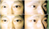
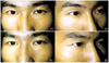
Most patients had a marked increase in orbital volume, as shown by the improvement of the upper eyelid sulcus deformity and enophthalmos (Fig. 1, 2, 3). All patients in this study tolerated well without intraoperative complications and there was no postoperative complications such as orbital hemorrhage, infection and infraorbital hypoesthesia. In all cases, postoperative pain was mild and easily controlled with analgesics. In no case extrusion or migration of the implant occurred during follow up period.
Discussion
Deep superior sulcus deformity and enophthalmos is a common problem in anophthalmic patients. Devoe4 described deepening of the superior sulcus in anophthalmic patients occurred due to malposition of the superior rectus muscle to the levator muscle that pulls the levator posteriorly instead of superiorly. In 1976, Vistnes1 added upperlid and lowerlid ptosis to enophthalmos with superior sulcus deformity and named "anophthalmic orbital syndrome". He suggested that chronic gravitational forces of the prosthesis might lead downward shift of the orbital contents and produced upperlid ptosis. In 1982, Tyers and Collin2 first described "postenucleation socket syndrome" composed of total appearance of enophthalmos, deepening of the upper eyelid sulcus, ptosis of the upper eyelid and stretching of the lower eyelid. Smit et al3 reviewed 22 cases of CT scan of the orbit and indicated different point of previously decribed postenucleation socket syndrome. The reviewed cases revealed retraction of upper eyelid in contrast to ptosis. Ptosis of the upper eyelid and fat atrophy was not observed, but rather, retraction of the superior muscle complex was noted to produce deepening of the sulcus and retraction of the upper eyelid. In our study, we noticed 2 cases of upper eyelid retraction and they needed recession of levator muscle.
Sometimes, prosthesis placement alone is often not adequate for the soft tissue loss in anophthalmic patients. The volume of an adult globe is approximately 8 cm3. Besides the 8 cm3 volume loss through enucleation, there can be additional soft tissue loss of 4 cm3, resulting in a total decreased volume of 12 cm3. The volume replaced by a 20 mm spherical implant approaches only 4 cm3. A prosthesis may give an additional 1.5 to 4 cm3, therefore providing a total 5.5 to 8 cm3 of orbital volume replacement, but still remain 4 to 6.5 cm3 deficit. If there is herniation of fat and displacement of the orbital wall, this discrepancy may be more prominent.16 In the case of evisceration, the severity of enophthalmos and superior sulcus deformity can be mild, but can also happen because of migration of orbital implant, atrophy of surrounding tissue and weight of implant.
The correction of this sunken eye appearance with upper and lower lid malposition has been a chief issue to oculoplastic surgeons, and many surgical techniques have been tried since Mules17 first attempted to replace the eviscerated eyeball with a artificial vitreous in 1885. Anophthalmic superior sulcus deformity can be addressed by a variety of techniques, including intraconal implants, subperiosteal implants or through direct augmentation of the superior sulcus. Direct augmentation of superior sulcus using free fat or dermis-fat graft, Alloderm®, temporoparietal fascia has been described for many years. However when using graft materials, resorption may happen with time and temporoparietal fascia flap surgery involve more complex surgery. Volume augmentation surgery for anophthalmic patients via subperiosteal pocket has several advantages over direct subcutaneous volume replacement. It may improve both enophthalmos and superior sulcus deformity, and may improve upper eyelid ptosis in certain cases. In enophthalmos correction, subperiosteal implant transplantation was effectively utilized for posttraumatic enophthalmos18,19 and anophthalmic enophthalmos.18 Replacement of lost volume has employed various materials, including glass beads,10 autologous or homologous bone,4 cartilage,11 and dermis,12 assorted plastic plate,13 room-temperature vulcanized(RTV) silicone,14 synthetic porous composite of Teflon polymer and alumina.15 The authors used porous polyethylene sheet already successfully used in orbital and craniofacial reconstruction during the past years. "Porous" or "integrated" implants are the most important graft used for orbital reconstructive surgery in recent years, with porous polyethylene and hydroxyapatite being the most common. Their porous structure allows for rapid vascular, soft-tissue and bone ingrowth that serves to stabilize the implant in relation to the surrounding tissue. Porous polyethylene is a highly biocompatible, durable and remarkably stable alloplast.20 Technically, it is easy to work, strong yet somewhat flexible and offers possibility of obtaining a precise three-dimensional shape.
In our study, superior sulcus deformity and enophthalmos was successfully corrected in most patients without complications. Two patients with retinoblastoma who received additional irradiation treatment in their childhood showed unsatisfactory cosmetic results (Grade 2). It is presumed that irradiation-induced slowing of bone growth can lead to a noticeable asymmetry in relative size and volume of the bony orbit and thus accentuating the appearance of enophthalmos.21 Furthermore, contraction or scarring of orbital soft tissue induced by irradiation can make the enophthalmos more prominent, so that the results of subperiosteal implantation of Medpor® sheets were not sufficient. There were 2 patients who showed improvement of upper eyelid ptosis without additional lid surgery nor exchanging their own prosthesis. We think that the volume deficit should be corrected first before upper eyelid ptosis surgery because an added orbital volume has some effects of pushing upward the upper eyelid.
In summary, subperiosteal implantation of porous high-density polyethylene sheet for anophthalmic patients with superior sulcus deformity and enophthalmos is shown to be a reliable and safe procedure with an excellent cosmetic results and without serious complications


 XML Download