

 PDF
PDF ePub
ePub Citation
 Print
Print


The mainstay of treatment for accommodative esotropia is a full correction of the hypermetropic refractive error, determined by cycloplegic refraction. However, esodeviation of refractive accommodative etiology does not always occur in a pure form. Residual esotropia may exist despite a full correction of hypermetropic refractive error. This non-accommodative component of esotropia should be surgically corrected. However, if the amount is about 10-15PD, we consider non-surgical treatment rather than surgery.
Hiatt1 described that miotics can be useful in supplemental glasses in a case of a small amount of residual esodeviation. Nevertheless, Miller2 cautioned that some patients experience gradually increased esotropia with prolonged miotics therapy. Furthermore, other side effects of miotics are well known.3
Occlusion therapy has proven to be the main tool of amblyopia treatment, and its risks are known to be minimal. However, full time occlusion might disrupt certain peripheral fusion which may control a latent component of the existing strabismus.4 Moreover, debate continues on whether occlusion may have an effect on preexisting esodeviation.5,6
We report two cases of suddenly deteriorated accommodative esotropia with mild amblyopia even during part-time occlusion therapy, and subsequently investigate the need of patching and the prognosis of such esotropia.
CASE REPORTS
Case 1
A 7-year-old girl presented to our department with an ingoing eye in her right eye.
She had worn glasses since 3 years of age. Her visual acuity was 20/25 in the right eye and 20/20 in the left. On initial examination, she had 2 PD of esophoria (E) for distant target and 10 PD esotropia for near target with the correction of refractive error. Without glasses she had 45 PD RET for distant target and 55 PD RET for near target. Cycloplegic refraction was performed and full spectacle correction was prescribed (right eye, +3.50 Diopters (DS); left eye, +2.50 DS). Visual acuity and ocular alignment were not changed with full spectacle correction. She had 800 sec of arc of stereoacuity confirmed with Titmus fly test with her new glasses. Her ocular examination was normal in other respects. She had no significant medical history. We prescribed new glasses and recommended patching the left eye for 3 hours a day for mild amblyopia in the right eye.
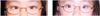
The corrected visual acuity in her right eye improved one line after 1 month, but she had 35 PD RET for distant and near targets with glasses (Fig. 1, left). The change of refraction did not occur during the periods of occlusion, as confirmed by repeated full cycloplegic refraction with atropinization. We therefore recommended surgery.
On preoperative evaluation after 1 month, alternate prism cover test showed orthophoria at distant target, and 14 PD RET at near target with glasses. Careful checking of history revealed that she had not actually undergone patching on her left eye during this period. We measured the angle of esodeviation after one-hour patching inducing manifestations of esotropia. After one hour, we noted that 35 PD RET was revealed at distant and near targets with glasses. We carefully observed her ocular alignment for several hours without patching, but the non-accommodative component of esotropia was unchanged. We performed 4.5 mm recession of both medial rectus muscles as scheduled. Orthophoria was well maintained at distant and near targets and she achieved fusion with stereoacuity of 60 seconds of arc until the last follow-up visit at 6 months after surgery (Fig. 1, right).
Case 2
A 9-year-old boy presented to our department with an ingoing eye in his right eye.
He had worn glasses and undergone part-time occlusion therapy from 5 years old. He had mild amblyopia in the right eye and his visual acuity was 20/25 in the right eye and 20/20 in the left. On initial examination, he had 35 PD RET for distant and near targets with glasses. Cycloplegic refraction was performed and full spectacle correction was prescribed (right eye, +3.00 DS; left eye, +3.00 DS). He had been wearing undercorrected glasses (right eye, +2.25, left eye, +2.00). The amount of esodeviation was decreased to 25 PD RET with a full spectacle correction. No binocularity was confirmed by Titmus fly test. The remainder of his ocular examination was normal. We prescribed new glasses and recommended patching the left eye for 3 hours a day.
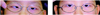
One month later, the amount of his ocular misalignment increased to 40 PD RET for distant and near targets with new glasses (Fig. 2, left). Full cycloplegic refraction was performed again, but no change of refraction was revealed during the periods of occlusion. We therefore recommended surgery.
On a preoperative evaluation 1 month later, alternate prism cover test showed orthophoria at distant target, and 12PD RET at near target with glasses (Fig. 2, right). He had never undergone patching on the left eye during this period. We cancelled surgery, and recommended wearing only glasses without patching.
Three months later, he gained 140 seconds of arc stereopsis. Orthophoria was maintained at distant target, and 8 PD esophoria at near target with his old glasses. We continued to observe the status of his ocular alignment for 6-months of follow-up.
DISCUSSION
Once amblyopia is diagnosed, occlusion treatment should be commenced without delay. Even preoperative occlusion therapy should be considered mandatory to achieve alternate fixation in congenital esotropia because the effectiveness of therapy depends directly on the age of the patient. However, treatment may still be successful even at the advanced age of 6 to 7 years and above. Provided compliance is good, part-time occlusion has proven to be an effective tool to maintain a good treatment result until the child reaches an age at which recurrence of amblyopia is no longer a problem, as occurred in our cases.7
It has been suggested that occlusion may have an effect on a preexisting esodeviation.5 However, Holbach et al6 described that actual occlusion did not cause the observed changes in ocular alignment of esotropic patients. However, they noted that the amount of changes in the deviation were significantly higher in the case of children under 42 months of age.
Besides the risk of occlusion amblyopia for the better eye, which is easily preventable with alternate patching and close monitoring, two patients reported by Swan8 had an underlying esophoria that became manifest during occlusion treatment for amblyopia when fusion was disrupted. Charney and Morris4 reported three esotropic children for whom full time occlusion precipitated an increased deviation of 10 to 43 PD. They described that the increase of the angle of deviation under these conditions is not an undesirable side-effect and that it may indicate exactly the amount of the deviation minus the effect of fusional amplitude. The decompensation of an angle may thus be the hallmark of good fusional potential result after surgery, similar to the "eat up" effect of the prism adaptation test.
The finding of a deteriorated preexisting esodeviation under occlusion must be distinguished from the acute comitant esotropia that occurs after artificial interruption of binocular vision, as it does after lid swelling, perforating corneal injury and cyclic esotropia, which have regular intervals but no insult like occlusion.
The well-controlled esophoria with glasses (case 1), and the preexisting small esotropia (case 2) of our patients might have been deteriorated and the angle of esodeviation increased due to part-time occlusion therapy. However, the binocularity of our patients was restored easily, with or without surgery, so we consider that the prognosis of such a deteriorated esotropia during occlusion therapy should be relatively excellent.
In conclusion, it is prudent to explain the risk of this complication to the parents or the patients before considering patching one eye. Furthermore, full cycloplegic refraction should be mandatory because an esotropia may ensue from wearing the patch in the presence of significantly uncorrected hypermetropic refractive error. We emphasize that occlusion therapy for esotropic patients who have amblyopia must be continuously taken, because an increase of the deviation size with occlusion may simply reflect a true deviation and may not be a poor prognostic sign.
 XML Download