

 PDF
PDF ePub
ePub Citation
 Print
Print


Abstract
Purpose
This study is executed to identify and report the treatment effects of oral mucosa grafting of prosthetic eye patients who have shallow conjunctival socket caused by socket contraction.
Methods
Conjunctival sac reconstruction was performed by using the lower lip mucosa to 11 eyes of 11 patients diagnosed with contracted conjunctival sac at the ophthalmic clinic from August 2009 to May 2015, and a retrospective analysis was performed with medical record data from the patients who were followed-up after surgery.
Results
Within the follow-up period, prosthetic eye insertion was possible for all 11 eyes of the 11 patients. All of them were satisfied in an aesthetic aspect, and were able to maintain deep conjunctival sac without receving findings of graft absorption, or re-contracted or shallow conjunctival sac during the follow-up period. On the donor region, normal epithelialization occurred. Concerning the donor region for the first week after surgery, the mean score of the patients' pain was 3.18 ± 0.94 points measured by a numerical rating scale, and no infection, bleeding, contraction, or cicatrix was revealed during the follow-up period. There were hypoesthesia, pararthria, and paresthesia (tingling sense) for post-complications from the donor region, and four patients out of the 11 patients complained of discomfort resulting from post-complications; whereas the remaining seven patients did not complain of discomfort.
REFERENCES
1). Soll DB. Reconstruction of a contracted socket. The use of an expandable silicone tire. Arch Ophthalmol. 1969; 82:218–9.
2). Guyuron B. Retroauricular island flap for eye socket reconstruction. Plast Reconstr Surg. 1985; 76:527–33.

3). Molgat YM, Hurtwitz JJ, Webb MC. Buccal mucous membrane-fat graft in the management of the contracted socket. Ophthal Plast Reconstr Surg. 1993; 9:267–72.
4). Suh IS, Yang YM, Oh SJ. Conjunctival cul-de-sac reconstruction with radial forearm free flap in anophthalmic orbit syndrome. Plast Reconstr Surg. 2001; 107:914–9.
5). Lee AC, Fedorovich I, Heinz GW, Kikkawa DO. Socket reconstruction with combined mucous membrane and hard palate mucosal grafts. Ophthalmic Surg Lasers. 2002; 33:463–8.
6). Sullivan SA, Dailey RA. Graft contraction: a comparison of acellular dermis versus hard palate mucosa in lower eyelid surgery. Ophthal Plast Reconstr Surg. 2003; 19:14–24.
7). Han JM, Choi HJ, Wee WR, et al. A case of alkali burn treated with amniotic membrane graft and forniceal reconstruction. J Korean Ophthalmol Soc. 2010; 51:1010–5.
8). Ballen PH. Mucous membrane grafts in chemical (lye) burns. Am J Ophthalmol. 1963; 55:302–12.
9). Lee SH, Kim JH, Kim JT, et al. A case of ocular surface reconstruction using nasal and oral mucosa autograft. J Korean Ophthalmol Soc. 2008; 49:1177–82.
10). Serin D, Karslıoğlu Ş, Akbaba M, et al. Clinical evaluation of 188 patients with contracted socket. Surgery Curr Res. 2014; 4:203.
11). Soll DB. The anophthalmic socket. Ophthalmology. 1982; 89:407–23.
12). Soll DB. Management of the anophthalmic socket and techniques of enucleation, evisceration, and exenteration: surgical procedures and management of complications. Tasman W, Jaeger EA, editors. Duane's Clinical Ophthalmology. revised ed.Philadelphia: JB Lippincott;1993. v. 5:chap. 83.
13). Lee TS, Hwang SJ, Oh JH. Forniceal reconstruction through subciliary approach in a patient with shallow inferior fornix. J Korean Ophthalmol Soc. 2007; 48:611–7.
14). Yang YH, Ahn M. Outcomes of autogenous dermis fat grafting with different donor sites in exposed porous orbital implants. J Korean Ophthalmol Soc. 2013; 54:545–51.
15). Yoon KC, Ji YS, Park YG. Management of exposed hydroxyapatite implant with acellular dermal allograft. J Korean Ophthalmol Soc. 2005; 46:927–32.
16). Rubin PA, Fay AM, Remulla HD, Maus M. Ophthalmic plastic applications of acellular dermal allografts. Ophthalmology. 1999; 106:2091–7.
17). Oh DE, Kim YD. Reconstruction of contracted anophthalmic socket with acellular dermal allograft. J Korean Ophthalmol Soc. 2008; 49:377–83.
18). Sclafani AP, Romo T 3rd, Jacono AA, et al. Evaluation of acellular dermal graft (AlloDerm) sheet for soft tissue augmentation: a 1-year follow-up of clinical observations and histological findings. Arch Facial Plast Surg. 2001; 3:101–3.
19). Kim JH, Chun YS, Lee SH, et al. Ocular surface reconstruction with autologous nasal mucosa in cicatricial ocular surface disease. Am J Ophthalmol. 2010; 149:45–53.
20). Kim YM, Son MG, Kim YD. Hard palate mucosa grafts for lower lid retraction. J Korean Ophthalmol Soc. 2000; 41:2319–26.
Figure 1.
Pre- and postoperative photo. (A) Postoperative photograph taken 11 days after the operation, conjunctival fornix reconstruction, upper. (B) Postoperative photograph taken 11 days after the operation, conjunctival fornix reconstruction, lower.
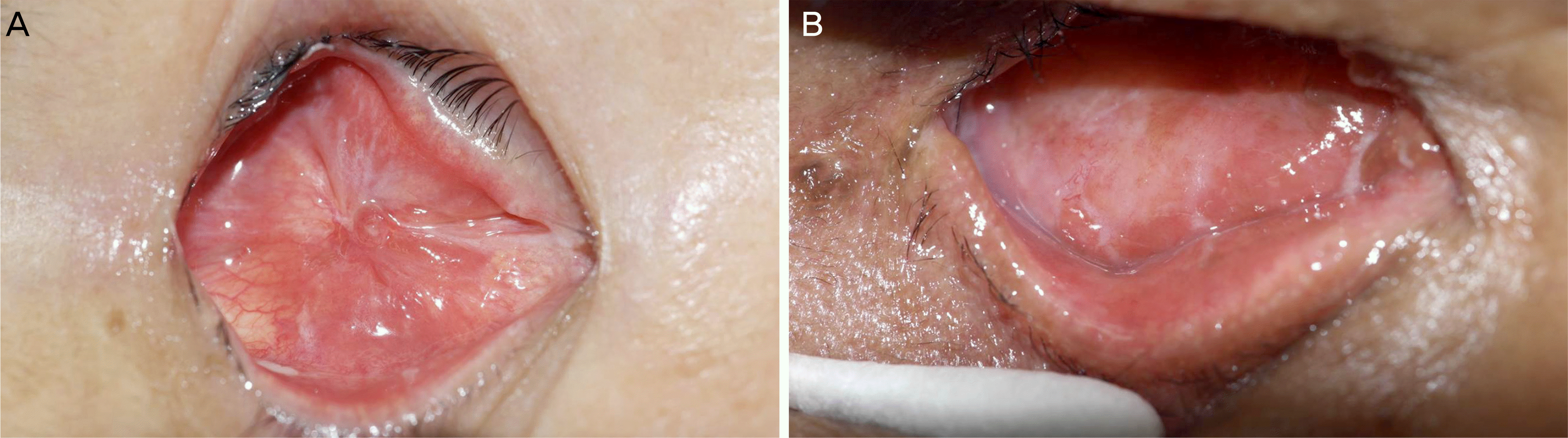
Figure 2.
Lower lip donor site. (A) Preoperative photograph. (B) Intraoperative photograph. (C) Postoperative photograph taken 4 days after the operation. (D) Postoperative photograph taken 11 days after the operation.

Table 1.
Grading of conjunctival socket contraction
Table 2.
Summary of patients
Table 3.
Summary of 11 operations, conjunctival sac reconstruction
Table 4.
Complications of donor site
| No. | Observation time (years) | NRS | Time to epithelization (weeks) | Answer* | Late phase complication |
|---|---|---|---|---|---|
| 1 | 1 | 3 | 4 | No | Hypoesthesia |
| 2 | 5 | 3 | 3 | Yes | - |
| 3 | 4 | 2 | 3 | Yes | Hypoesthesia |
| 4 | 5 | 2 | 2 | Yes | - |
| 5 | 5 | 5 | 3 | No | - |
| 6 | 5 | 3 | 3 | Yes | Hypoesthesia, tingling sensation |
| 7 | 5 | 3 | 2 | Yes | - |
| 8 | 5 | 3 | 3 | Yes | - |
| 9 | 4 | 3 | 3 | Yes | - |
| 10 | 1 | 3 | 3 | Yes | - |
| 11 | 2/12 | 5 | 5 | No | Hypoesthesia, numbness |
 XML Download