

 PDF
PDF ePub
ePub Citation
 Print
Print


Abstract
Purpose
To investigate the postural change of intraocular pressure (IOP) from sitting to supine position and determine the relationship to other ocular parameters including ocular pulse amplitude (OPA) in glaucoma suspect and open angle glaucoma patients.
Methods
The present study included 46 eyes of 46 patients. First, we measured IOP and OPA using Goldmann applanation tonometer (GAT), Pascal dynamic contour tonometer and TonoPen®. Using TonoPen®, the IOP was measured immediately after the subjects were placed in a supine position and 10 minutes and 30 minutes thereafter. We also investigated the correlation between positional change of IOP and axial length (AL), refractive error (RE), and OPA.
Results
IOPs of patients in a sitting position measured with GAT and TonoPen® were 15.3 ± 3.3 mm Hg and 16.6 ± 2.9 mm Hg, respectively, and OPA was 2.57 ± 0.89 mm Hg. IOPs measured with TonoPen® were 17.6 ± 2.9 mm Hg immediately after position change, 18.2 ± 3.7 mm Hg after 10 minutes and 17.5 ± 2.7 mm Hg after 30 minutes. Each IOP change was statistically significant and the largest change was after 10 minutes. Changes of IOP after 10 minutes were positively correlated with OPA (R = 0.340) and RE (R = 0.330) and negatively correlated with AL (R = -0.410).
References
1. Allingham RR, Damji KF. Shields textbook of glaucoma. 6th ed.Philadelphia: Wolters Kluwer Health/Lippincott Williams & Wilkins;2011. chap. 2.
2. Leonard TJ, Kerr Muir MG, Kirkby GR, Hitchings RA. Ocular hypertension and posture. Br J Ophthalmol. 1983; 67:362–6.

3. Tsukahara S, Sasaki T. Postural change of IOP in normal persons and in patients with primary wide open-angle glaucoma and low-tension glaucoma. Br J Ophthalmol. 1984; 68:389–92.
4. Carlson KH, McLaren JW, Topper JE, Brubaker RF. Effect of body position on intraocular pressure and aqueous flow. Invest Ophthalmol Vis Sci. 1987; 28:1346–52.
5. Lee JY, Yoo C, Kim YY. The effect of lateral decubitus position on intraocular pressure in patients with untreated open-angle glaucoma. Am J Ophthalmol. 2013; 155:329–35.e2.
6. Heijl A, Leske MC, Bengtsson B. . Reduction of intraocular pressure and glaucoma progression: results from the Early Manifest Glaucoma Trial. Arch Ophthalmol. 2002; 120:1268–79.
7. Jonas JB, Budde WM, Stroux A. . Diurnal intraocular pressure profiles and progression of chronic open-angle glaucoma. Eye (Lond). 2007; 21:948–51.
8. Dumskyj MJ, Mathias CJ, Doré CJ. . Postural variation in in-traocular pressure in primary chronic autonomic failure. J Neurol. 2002; 249:712–8.
9. Grieshaber MC, Katamay R, Gugleta K. . Relationship between ocular pulse amplitude and systemic blood pressure measurements. Acta Ophthalmol. 2009; 87:329–34.
10. Dastiridou AI, Ginis HS, De Brouwere D. . Ocular rigidity, ocular pulse amplitude, and pulsatile ocular blood flow: the effect of intraocular pressure. Invest Ophthalmol Vis Sci. 2009; 50:5718–22.
11. Stalmans I, Harris A, Vanbellinghen V. . Ocular pulse amplitude in normal tension and primary open angle glaucoma. J Glaucoma. 2008; 17:403–7.
12. Lee M, Cho EH, Lew HM, Ahn J. Relationship between ocular pulse amplitude and glaucomatous central visual field defect in normal-tension glaucoma. J Glaucoma. 2012; 21:596–600.
13. Araie M, Sekine M, Suzuki Y, Koseki N. Factors contributing to the progression of visual field damage in eyes with normal-tension glaucoma. Ophthalmology. 1994; 101:1440–4.
14. Daugeliene L, Yamamoto T, Kitazawa Y. Risk factors for visual field damage progression in normal-tension glaucoma eyes. Graefes Arch Clin Exp Ophthalmol. 1999; 237:105–8.
15. Grieshaber MC, Flammer J. Blood flow in glaucoma. Curr Opin Ophthalmol. 2005; 16:79–83.
16. Harris A, Rechtman E, Siesky B. . The role of optic nerve blood flow in the pathogenesis of glaucoma. Ophthalmol Clin North Am. 2005; 18:345–53. v.
17. Flammer J, Orgül S, Costa VP. . The impact of ocular blood flow in glaucoma. Prog Retin Eye Res. 2002; 21:359–93.
18. Galassi F, Giambene B, Varriale R. Systemic vascular dysregulation and retrobulbar hemodynamics in normal-tension glaucoma. Invest Ophthalmol Vis Sci. 2011; 52:4467–71.
19. Jain MR, Marmion VJ. Rapid pneumatic and Mackey-Marg appla-nation tonometry to evaluate the postural effect on intraocular pressure. Br J Ophthalmol. 1976; 60:687–93.
20. Krieglstein GK, Waller WK, Leydhecker W. The vascular basis of the positional influence of the intraocular pressure. Albrecht Von Graefes Arch Klin Exp Ophthalmol. 1978; 206:99–106.
21. Hvidberg A, Kessing SV, Fernandes A. Effect of changes in PCO2 and body positions on intraocular pressure during general anaesthesia. Acta Ophthalmol (Copenh). 1981; 59:465–75.
22. Sawada A, Yamamoto T. Posture-induced intraocular pressure changes in eyes with open-angle glaucoma, primary angle closure with or without glaucoma medications, and control eyes. Invest Ophthalmol Vis Sci. 2012; 53:7631–5.
23. Kiuchi T, Motoyama Y, Oshika T. Postural response of intraocular pressure and visual field damage in patients with untreated nor-mal-tension glaucoma. J Glaucoma. 2010; 19:191–3.
24. Kiuchi T, Motoyama Y, Oshika T. Relationship of progression of visual field damage to postural changes in intraocular pressure in patients with normal-tension glaucoma. Ophthalmology. 2006; 113:2150–5.
25. Fontana L, Poinoosawmy D, Bunce CV. . Pulsatile ocular blood flow investigation in asymmetric normal tension glaucoma and normal subjects. Br J Ophthalmol. 1998; 82:731–6.
26. Park SC, De Moraes CG, Teng CC. . Initial parafoveal versus peripheral scotomas in glaucoma: risk factors and visual field characteristics. Ophthalmology. 2011; 118:1782–9.
27. Lam AK, Chan ST, Chan B, Chan H. The effect of axial length on ocular blood flow assessment in anisometropes. Ophthalmic Physiol Opt. 2003; 23:315–20.
28. Dastiridou AI, Ginis H, Tsilimbaris M. . Ocular rigidity, ocular pulse amplitude, and pulsatile ocular blood flow: the effect of axial length. Invest Ophthalmol Vis Sci. 2013; 54:2087–92.
29. Jang SR, Lee MV, Ahn JH. Comparison of dorzolamide-timolol fixed combination and latanoprost, effects on intraocular pressure and ocular pulse amplitude. J Korean Ophthalmol Soc. 2014; 55:854–9.
30. Lu LC, Wei TM, Li S. . Differences in blood pressure readings between supine and sitting positions in hypertensive patients. Acta Cardiol. 2008; 63:707–11.
Figure 1.
Illustration of postural change of our subjects. (A) Sitting in-traocular pressure (IOP) was measure while subjects sitting on chair vertically. (B) Supine IOP was measured while subjects lying on the bed without a pillow.
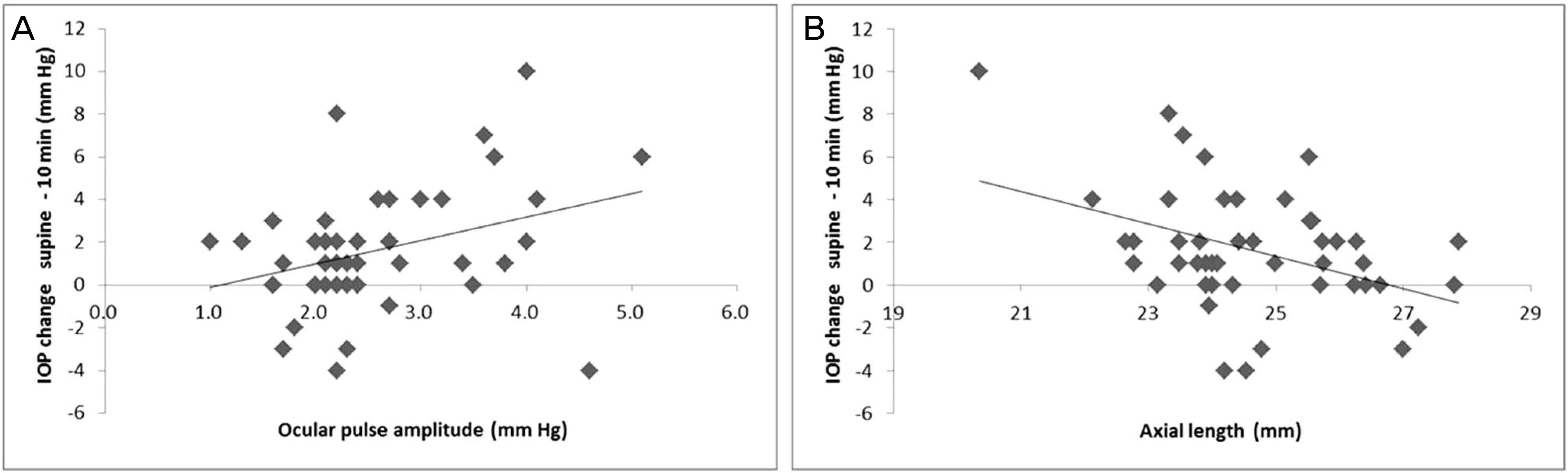
Figure 2. ( A) Correlation between ocular pulse amplitude and change of intraocular pressure (IOP) between sitting IOP and supine IOP 10 minutes after taking supine position. (B) Correlation between axial length and changes of IOP between sitting IOP and supine IOP 10 minutes after taking supine position.
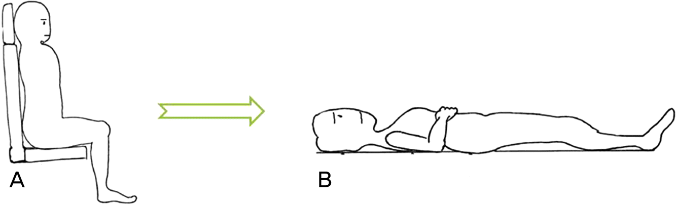
Table 1.
Descriptive statistics of 46 subjects
Table 2.
IOP and OPA measured at each time point
| Sitting | Supine-0 minute | Supine-10 minutes | Supine-30 minutes | |
|---|---|---|---|---|
| GAT IOP (mm Hg) | 15.3 ± 3.3 | - | - | - |
| DCT IOP (mm Hg) | 18.8 ± 4.1 | - | - | - |
| DCT OPA (mm Hg) | 2.6 ± 0.8 | - | - | - |
| TonoPen® IOP (mm Hg) | 16.6 ± 2.9 | 17.1 ± 2.9∗ | 18.2 ± 3.7∗ | 17.5 ± 2.8∗ |
Table 3.
Correlation between positional IOP change and ocular parameters
Table 4.
Correlation between positional IOP change and severity of glaucomatous visual field defects
 XML Download