

 PDF
PDF ePub
ePub Citation
 Print
Print


Abstract
Purpose
We compared ocular torsion rates in blowout fracture patients before and after blowout fracture repair by analyzing mean disc foveal angles.
Methods
The study participants were divided into 2 groups: blowout fracutre repair patients (n = 36) and controls (n = 36). We measured ocular torsion rates by analyzing mean disc foveal angle. The angle was composed of 2 imaginary horizontal lines which crossed the optic disc center and fovea. We compared statistically ocular torsion rates in blow-out fracture patients based on subsided diplopia, continued diplopia, or absence of diplopia before and after blow-out fracture repair using paired t-test.
Results
In the patient group, ocular torsion rates were statistically significantly decreased. In the blow-out fracture repair group with subsided diplopia, ocular torsion rates were decreased statistically from 7.74 ± 3.48 degrees before blow-out fracture repair to 5.02 ± 3.11 degrees after blow-out fracture repair. In the blow-out fracture repair group with continued diplopia or absence of diplopia before surgery, ocular torsion rates did not change statistically significantly from 6.36 ± 2.80 degrees before blow-out fracture repair to 6.51 ± 3.24 degrees after blow-out fracture repair.
Conclusions
Subsided diplopia after blow-out fracture repair and ocular torsion rate changes were significantly related in blow-out fracture patients. Further research which on the correlation of intraorbital change and movement of orbital position after blow-out fracture repair with ocular torsion rates are necessary.
References
1. Kim SK, Chang HK. The clinical study of treatment of blowout fracture. J Korean Ophthalmol Soc. 1995; 36:1629–35.
2. Kim HE, Lew H, Yun YS. The size of extraocular muscles estimated by computed tomography in patients undergoing orbital wall fracture repair. J Korean Ophthalmol Soc. 2009; 50:1447–54.

3. Poeschl PW, Baumann A, Dorner G. . Functional outcome after surgical treatment of orbital floor fractures. Clin Oral Investig. 2012; 16:1297–303.
4. Thiagarajah C, Kersten RC. Medial wall fracture: an update. Craniomaxillofac Trauma Reconstr. 2009; 2:135–9.
5. Hwang JH, Kwak MS. Residual Diplopia and Enophthalmos after Reconstruction of Orbital Wall Fractures. J Korean Ophthalmol Soc. 2003; 44:1959–65.
6. Cha MB, Min BM, Choi SH. Analysis of ocular motility disturbance remained after open reduction in orbital wall fracture. J Korean Ophthalmol Soc. 1997; 38:1885–91.
7. Ceylan OM, Uysal Y, Mutlu FM. . Management of diplopia in patients with blowout fractures. Indian J Ophthalmol. 2011; 59:461–4.
8. Joseph JM, Glavas IP. Orbital fractures: a review. Clinical Ophthalmol. 2011; 5:95.
9. Kim HW, Kim YI, Won IK. Clinical analysis of blowout fracture with ocualr motion limitation: comparison of surgical and conservative treatment. J Korean Ophthalmol Soc. 1999; 40:632–8.
10. Kushner BJ, Hariharan L. Observations about objective and subjective ocular torsion. Ophthalmology. 2009; 116:2001–10.
11. Lee HJ, Lim KH. The range of ocular torsion in mass screening. J Korean Ophthalmol Soc. 2005; 46:1684–9.
12. Kothari MT, Venkatesan G, Shah JP. . Can ocular torsion be measured using the slitlamp biomicroscope? Indian J Ophthalmol. 2005; 53:43–7.
13. Kim EH, Lee SJ, Choi HY. Ocular torsion according to fixation in fundus photograph. J Korean Ophthalmol Soc. 2006; 47:449–54.
14. Bixenman WW, von Noorden GK. Apparent foveal displacement in normal subjects and in cyclotropia. Ophthalmology. 1982; 89:58–62.
15. Park KH, Shin JH, Kim SY. Surgical results of modified Harada-Ito operation for excyclotorsion. J Korean Ophthalmol Soc. 2012; 53:565–71.
16. Kang HJ, Ha MS. A clinical feature of the patients of orbital wall fracture with diplopia. J Korean Ophthalmol Soc. 2009; 50:969–75.
17. Pearl RM. Treatment of enophthalmos. Clin Plast Surg. 1992; 19:99–111.
18. Iliff NT. The ophthalmic implications of the correction of late enophthalmos following severe midfacial trauma. Trans Am Ophthalmol Soc. 1991; 89:477–548.
19. Converse JM, Smith B, Obear MF, Wood-Smith D. Orbital blow-out fractures: a ten-year survey. Plast Reconstr Surg. 1967; 39:20–36.
20. Burres SA, Cohn AM, Mathog RH. Repair of orbital blowout fractures with Marlex mesh and Gelfilm. Laryngoscope. 1981; 91:1881–6.
21. Lee SJ, Park KS. Relationship between preoperative clinical features and postoperative recovery of ocular motility restriction in blow-out fractures. J Korean Ophthalmol Soc. 2001; 42:1202–9.
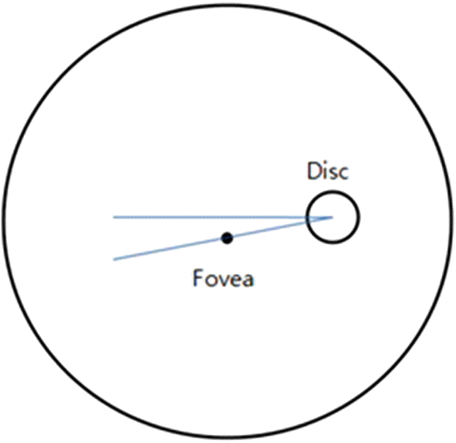
Figure 2.
1: The patients who had blow out fracture of the inferior wall complained of moderate degree diplopia. Before the operation, 12.7 degree of disc foveal angle was observed on fundus photographs. Almost 10 degrees of extorsion was checked with double moddox rod test. Six months after the operation, the patients did not complain of diplopia. 4.3 degree of disc foveal angle was observed on fundus photographs No extorstion and intorsion were checked with double moddox rod test. 2: The patients who had blow out fracture of the inferior wall complained of moderate degree diplopia. Before the operation, 11.2 degree of disc foveal angle was observed on fundus photographs. Almost 6 degrees of extorsion was checked with double moddox rod test. Six months after the operation, the patients did not complain of diplopia. 6.2 degree of disc foveal angle was observed at the fundus photograph. No extorstion and intorsion were checked with double moddox rod test. 3: The patients who had blow out fracture of the inferior wall complained of moderate degree diplopia. Before the operation, 9.6 degree of disc foveal angle was observed on fundus photographs. Almost 5 degree of extorsion was checked with double moddox rod test. 6 months after the operation, the patients did not complain of diplopia. 4.1 degree of disc foveal angle was observed at the fundus photograph. No extorstion and intorsion were checked with double moddox rod test.
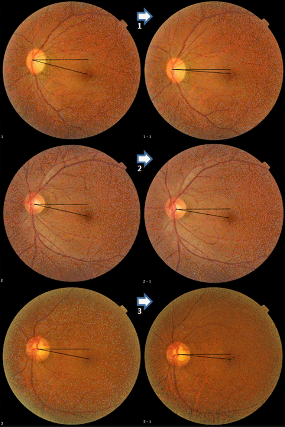
Table 1.
Baseline characteristics of blow out fracture patients
| Factors | Values |
|---|---|
| Total eyes (n) | 36 |
| Age (years) | 34.6 ± 15.7 (12-70) |
| Sex | |
| Male | 30 |
| Female | 6 |
| Laterality | |
| Right | 15 |
| Left | 21 |
| Duration of F/U (months) | 9.4 ± 2.7 |
| Operation period (days) | 6.7 ± 1.2 |
Table 2.
Location of blow out fracture
| Involved wall | Number of cases |
|---|---|
| Inferior wall | 13 |
| Medial wall | 15 |
| Inferior wall + lateral wall | 0 |
|
Inferior wall + medial wall |
8 |
| Total | 36 |
Table 3.
Muscle status of blow out fracture patients
| Muscle status | Number of cases |
|---|---|
| Herniation | 6 |
| Incaceration | 2 |
| Thickening | 9 |
| Thickening & herniation | 15 |
|
None |
4 |
| Total | 36 |
Table 4.
Diplopia of blow out fracture patients
| Diplopia | Number of cases |
|---|---|
| Absent | 15 |
| Mild | 6 |
| Moderate | 14 |
|
Severe |
1 |
| Total | 36 |
Table 5.
Ocular torsion rate in blow out fracture patient group & control group
|
Blow out fracture group |
Control group | ||
|---|---|---|---|
| Pre-operation | Post-operation | ||
| Ocular torsion rate (degree) | 7.07 ± 5.71 | 5.71 ± 3.24 | 5.88 ± 3.34 |
 XML Download