

 PDF
PDF ePub
ePub Citation
 Print
Print


Abstract
Purpose
We compared the measurements of anterior chamber depth (ACD) and anterior chamber angle (ACA) using ultrasound biomicroscopy (UBM) in the sitting position compared with IOL Master®, Pentacam®, and Spectralis optical coherence tomography (OCT) to evaluate the clinical usefulness of UBM in the sitting position.
Methods
We evaluated 92 eyes in 47 healthy adults. ACD was measured by IOL Master®, Pentacam®, and UBM. ACA was measured using Pentacam®, UBM, and Spectralis OCT. UBM was performed in the sitting position using bag/balloon technology. Measured values were compared statistically.
Results
ACD measured by IOL Master®, Pentacam®, and UBM was 3.57 ± 0.32 μm, 3.64 ± 0.33 μm and 3.51 ± 0.32 μm, respectively. UBM measurements of ACD were significantly shallower than with the other methods (p < 0.001). The results among the 3 methods were strongly correlated (r > 0.8, p < 0.05 in all groups). Mean ACA of 4 directions was not significantly different among the 3 methods (p > 0.05). There was strong correlation between UBM and Spectralis OCT (r = 0.957) but moderate correlation between Pentacam® and UBM and Pentacam® and Spectralis OCT (r = 0.557, 0.571, respectively, p < 0.05). Specifically, ACA of the superior quadrant showed a low correlation between Pentacam® and UBM and Pentacam® and Spectralis OCT (r = 0.257, 0.295, respectively).
Conclusions
ACD measured by UBM in the sitting position was shallower compared to the other methods; however, ACD measured by IOL Master®, Pentacam®, and UBM showed significant correlations among the methods. The mean ACA measured by Pentacam®, UBM, and Spectralis OCT showed no significant differences. Due to the high correlation of ACA measurements between UBM and Spectralis OCT in the present study, UBM is expected to be a good tool for measuring anterior segment parameters.
References
1. Olsen T. Sources of error in intraocular lens power calculation. J Cataract Refract Surg. 1992; 18:125–9.

2. Holladay JT. Standardizing constants for ultrasonic biometry, keratometry, and intraocular lens power calculations. J Cataract Refract Surg. 1997; 23:1356–70.
3. Alio JL, de la Hoz F, Perez-Santonja JJ, et al. Phakic anterior chamber lenses for correction of myopia; a 7-year cumulative analysis of complications in 263 cases. Ophthalmology. 1999; 106:458–66.
4. Holladay JT. Refractive power calculations for intraocular lenses in the phakic eye. Am J Ophthalmol. 1993; 116:63–6.
5. Kashiwagi K, Tokunaga T, Iwase A, et al. Agreement between peripheral anterior chamber depth evaluation using the van Herick technique and angle width evaluation using the Shaffer system in Japanese. Jpn J Ophthalmol. 2005; 49:134–6.
6. Narayanaswamy A, Vijaya L, Shantha B, et al. Anterior chamber angle assessment using gonioscopy and ultrasound biomicroscopy. Jpn J Ophthalmol. 2004; 48:44–9.
7. Koranyi G, Lydahl E, Norrby S, Taube M. Anterior chamber depth measurement: a-scan versus optical methods. J Cataract Refract Surg. 2002; 28:243–7.
8. Konstantopoulos A, Hossain P, Anderson DF. Recent advances in ophthalmic anterior segment imaging: a new era for ophthalmic diagnosis. Br J Ophthalmol. 2007; 91:551–7.
9. Sakata LM, Wong TT, Wong HT, et al. Comparison of Visante and slit-lamp anterior segment optical coherence tomography in imaging the anterior chamber angle. Eye (Lond). 2010; 24:578–87.
10. Nasser CK, Singer R, Barkana Y, et al. Repeatability of the Sirius imaging system and agreement with the Pentacam HR. J Refract Surg. 2012; 28:493–7.
11. Jea SY, Jung SC, Oum BS. Quantified values of anterior chamber depth and angle measurements using ultrasound biomicroscopy and topography. J Korean Ophthalmol Soc. 2006; 47:97–104.
12. Park YS, Ahn H, Kim NR, et al. Comparison of measurement of anterior segment parameters between scheimpflug camera and ultrasound biomicroscopy. J Korean Ophthalmol Soc. 2009; 50:1847–52.
13. Dada T, Sihota R, Gadia R, et al. Comparison of anterior segment optical coherence tomography and ultrasound biomicroscopy for assessment of the anterior segment. J Cataract Refract Surg. 2007; 33:837–40.
14. He M, Foster PJ, Ge J, et al. Gonioscopy in adult Chinese: the Liwan eye study. Invest Ophthalmol Vis Sci. 2006; 47:4772–9.
15. Esaki K, Ito K, Matsunaga K, et al. Anterior chamber structural change in postural variation in pseudoexfoliation syndrome. Nihon Ganka Gakkai Zasshi. 2001; 105:524–9.
16. Bell NP, Nagi KS, Cumba RJ, et al. Age and positional effect on the anterior chamber angle: assessment by ultrasound biomicroscopy. ISRN Ophthalmology. 2013; 2013:706201.
17. Bell NP, Feldman RM, Zou Y, Prager TC. New technology for examining the anterior segment by ultrasonic biomicroscopy. J Cataract Refract Surg. 2008; 34:121–5.
18. Kiraly L, Duncker G. Biometry of the anterior eye segment for implantation of phakic anterior chamber lenses. A comparison of current measurement devices. Ophthalmologe. 2012; 109:242–9.
19. Holladay JT, Prager TC, Chandler TY, et al. A three-part system for refining intraocular lens power calculations. J Cataract Refract Surg. 1988; 14:17–24.
20. Liang J, Liu W, Xing X, et al. Evaluation of the agreement between Pentacam and ultrasound biomicroscopy measurements of anterior chamber depth in Chinese patients with primary angle-closure glaucoma. Jpn J Ophthalmol. 2010; 54:361–2.
21. Lee DG, Choi SH. Measurement of anterior segment using visante OCT in Koreans. J Korean Ophthalmol Soc. 2009; 50:542–50.
22. Oh DE, Jun RM, Choi KR. Quantified values of anterior segment in normal adult Koreans using ultrasound biomicroscopy. J Korean Ophthalmol Soc. 2004; 45:251–8.
23. Reuland MS, Reuland AJ, Nishi Y, Aufarth GU. Corneal radii and anterior chamber depth measurements using the IOLmaster versus the Pentacam. J Refract Surg. 2007; 23:368–73.
24. Elbaz U, Barkana Y, Gerber Y, et al. Comparison of different techniques of anterior chamber depth and keratometric measurements. Am J Ophthalmol. 2007; 143:48–53.
25. Philips CI. Closed-angle glaucoma; significance of sectoral variations in angle depth. Br J Ophthalmol. 1956; 40:136–43.
26. Böker T, Sheqem J, Rauwolf M, Wegener A. Anterior chamber angle biometry: a comparison of Scheimpflug photography and ultrasound biomicroscopy. Ophthalmic Res. 1995; 27:104–9.
27. Yi JH, Hong S, Seong GJ, et al. Anterior chamber measurements by pentacam and AS-OCT in eyes with normal open angles. Korean J Ophthalmol. 2008; 22:242–5.
Figure 1.
An example of the result of anterior chamber angle (ACA) measurement by Pentacam®. (A) Nasal and temporal angle. (B) Superior and inferior angle. ACA is calculated by integrated software of Pentacam®.
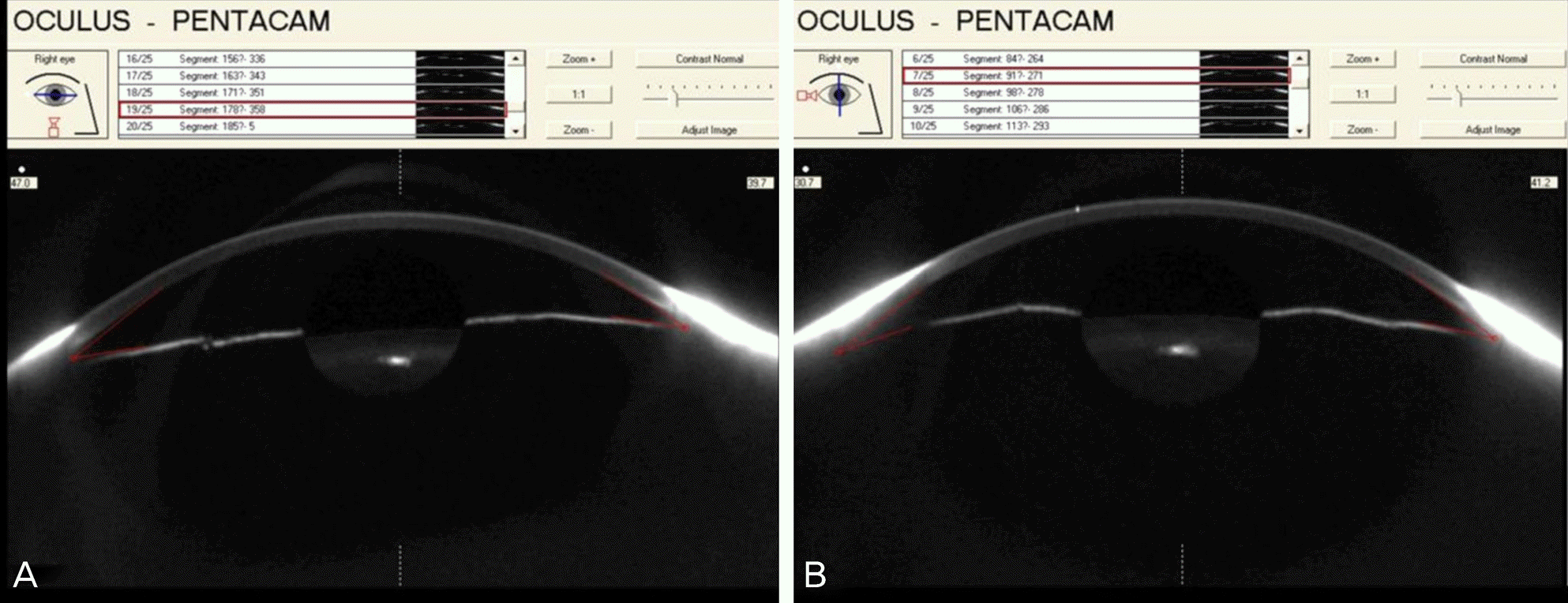
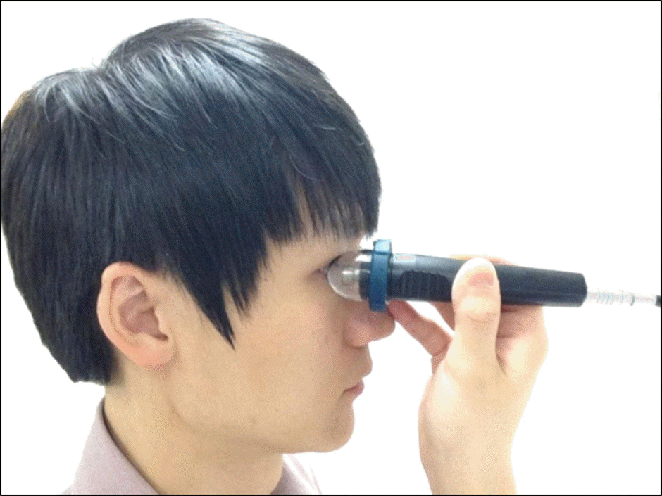
Figure 3.
Ultrasound biomicroscopic image of anterior chamber and lens. Anterior chamber depth 1 (ACD1) is defined as distance from the central corneal epithelium to the anterior surface of lens capsule and ACD2 is defined as distance from the central corneal endothelium to the anterior surface of lens capsule.
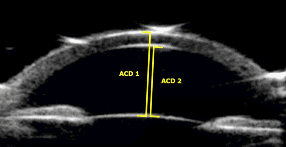
Figure 4.
Ultrasound biomicroscopic (A) and spectralis optical coherence tomography (OCT), (B) image of anterior chamber angle. Anterior chamber angle (ACA) is measured by trabecular iris angle (TIA). TIA is defined as the angle between the iris and trabecular meshwork, with the apex of the angle lying in the angle recess and arms lying at a point 500 μm away from the scleral spur.
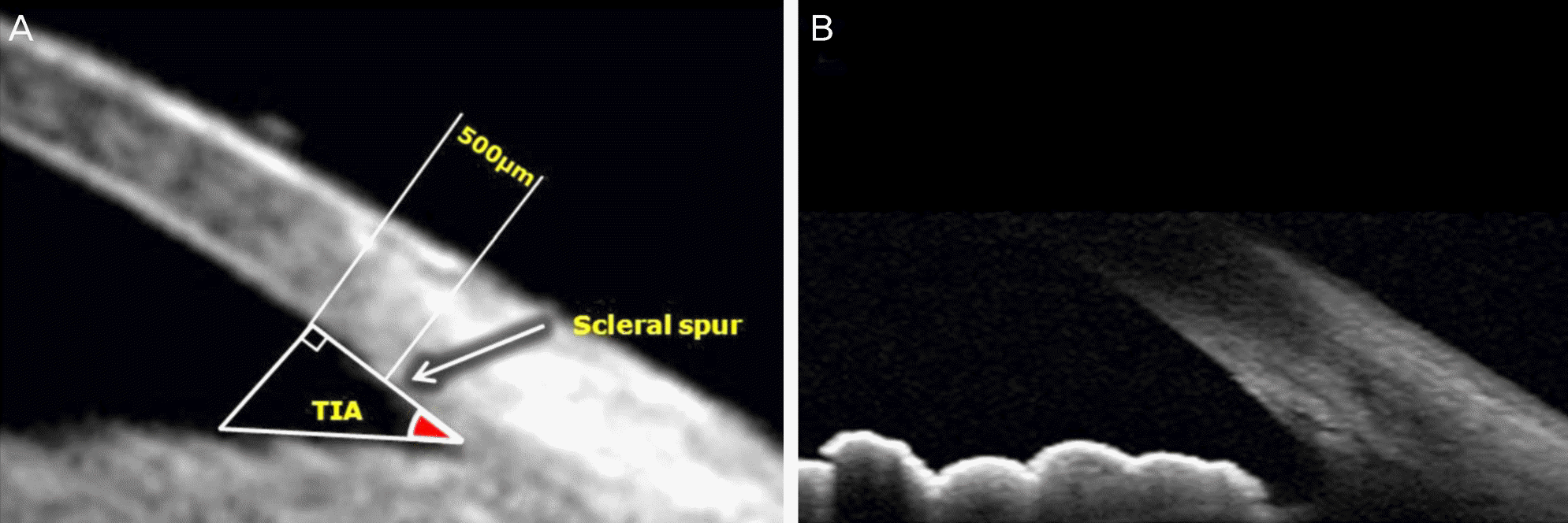
Figure 5.
Bland-Altman plots showing differences in anterior chamber depth (ACD) between devices. The upper and lower dashed lines indicate 95 % limits of agreement and the solid line indicates the mean. UBM = ultrasound biomicroscopy.
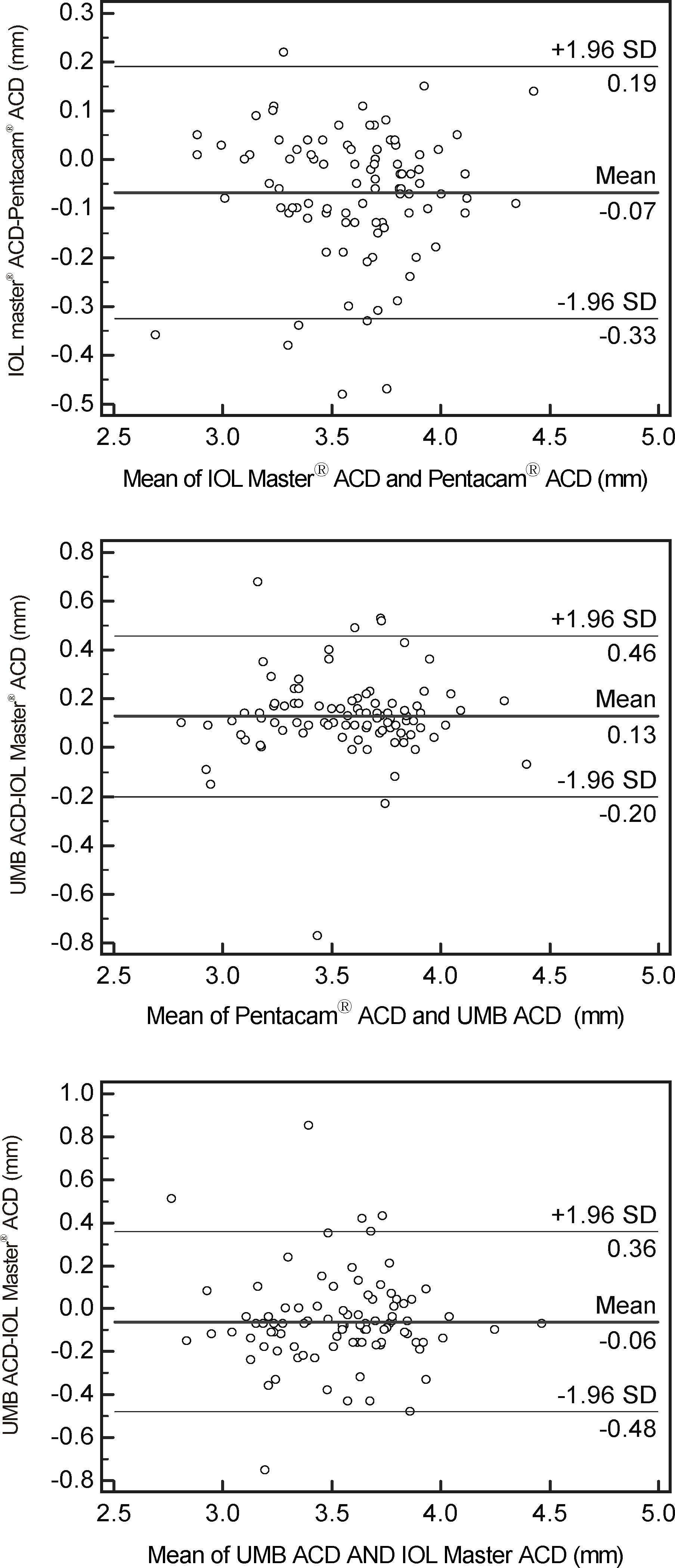
Figure 6.
The correlation plot of the anterior chamber depth (ACD) measured by IOL master®, Pentacam® and ultrasound biomicroscopy (UBM).
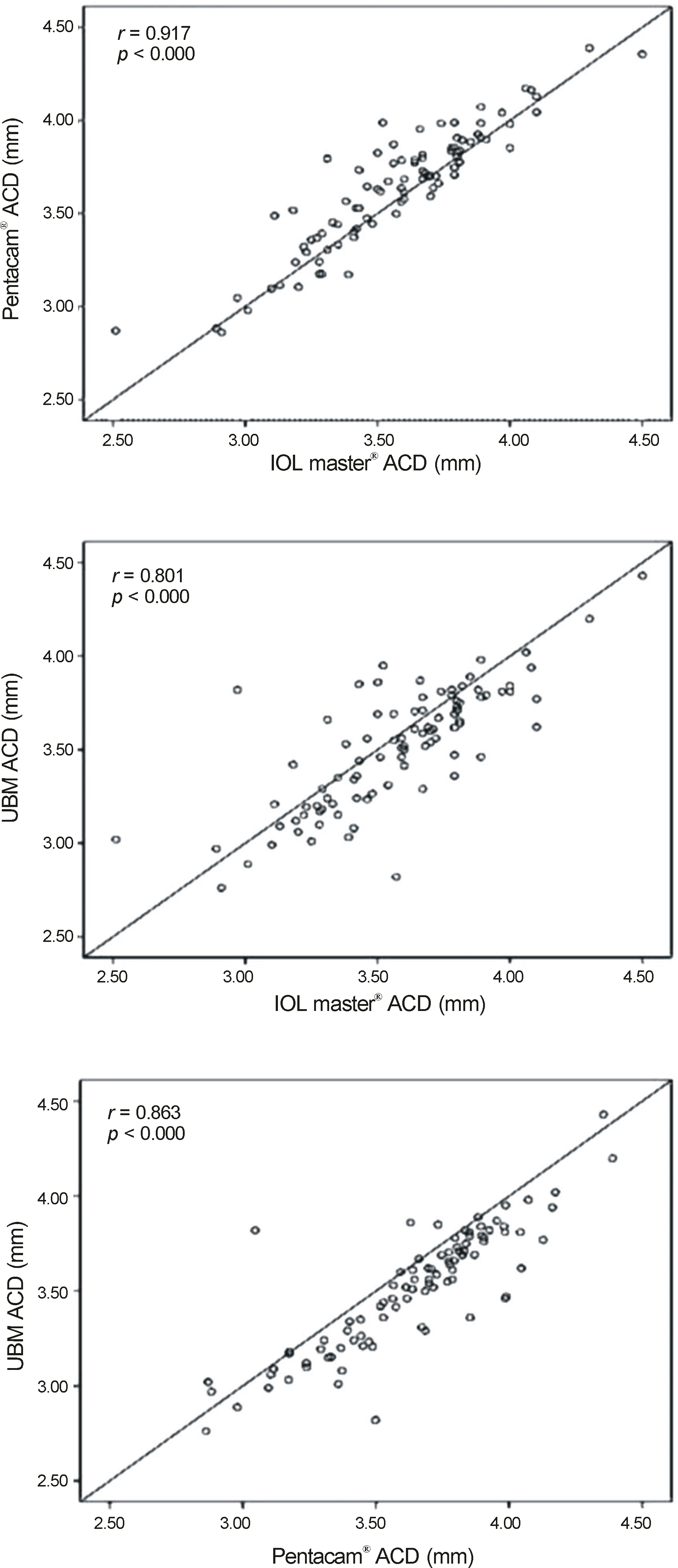
Figure 7.
Bland-Altman plots showing differences in anterior chamber angle (ACA) between devices. The upper and lower dashed lines indicate 95 % limits of agreement and the solid line indicates the mean. OCT = optical coherence tomography; UBM = ultrasound biomicroscopy.
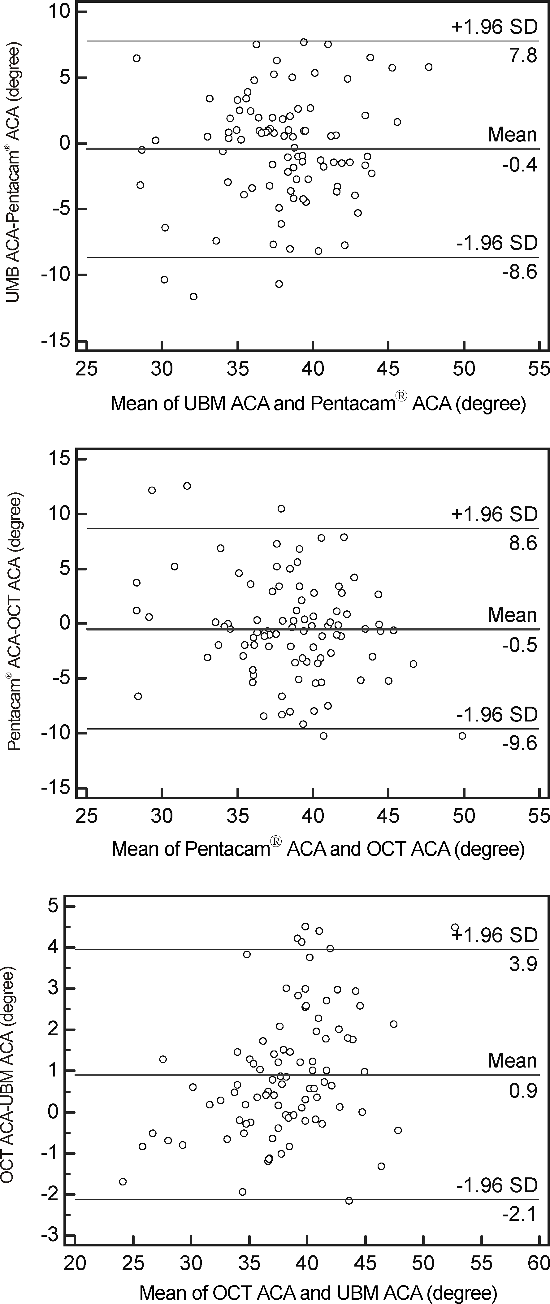
Figure 8.
The correlation plot of the mean anterior chamber angle (ACA) measured by Pentacam®, Spectralis OCT and ultrasound biomicroscopy (UBM). OCT = optical coherence tomography.
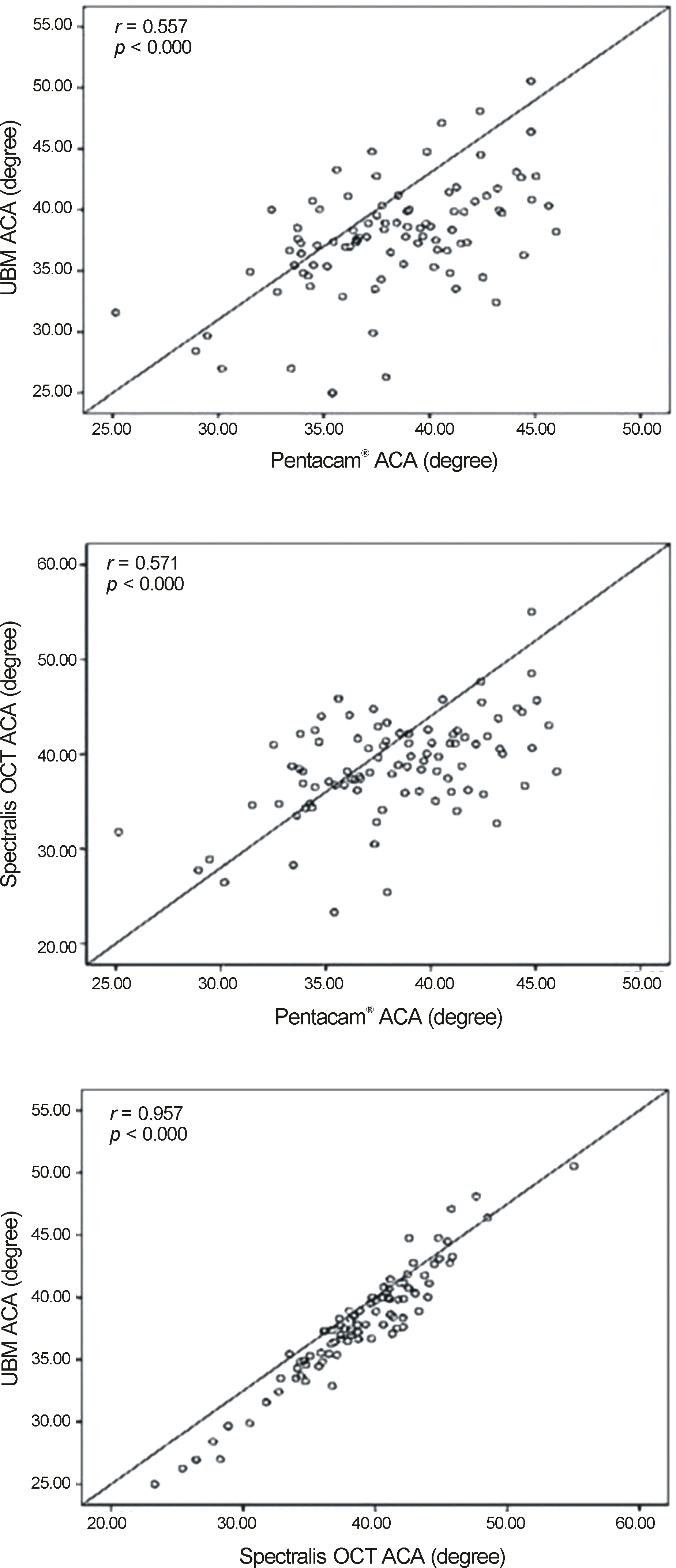
Table 1.
Comparison of different techniques of anterior chamber depth (ACD)
| IOL master® | Pentacam® | UBM | p-value* | ||
|---|---|---|---|---|---|
| ACD1 | Male | 3.60 ± 0.36 | 3.66 ± 0.36 | 3.52 ± 0.36 | <0.001 |
| Female | 3.51 ± 0.20 | 3.59 ± 0.23 | 3.48 ± 0.25 | <0.001 | |
| Mean | 3.57 ± 0.32 | 3.64 ± 0.33 | 3.51 ± 0.32 | <0.001 | |
| ACD2 | Male | NA | 3.10 ± 0.36 | 2.95 ± 0.25 | <0.001 |
| Female | NA | 3.04 ± 0.23 | 2.94 ± 0.35 | <0.001 | |
| Mean | NA | 3.08 ± 0.33 | 2.95 ± 0.33 | <0.001 |
Table 2.
Anterior chamber angle (ACA) at different directions and mean ACA
| Directions |
Anterior chamber angle (degree) |
|||
|---|---|---|---|---|
| Pentacam® | UBM | Spectralis-OCT | ||
| Temporal ACA | Male | 41.78 ± 6.10 | 40.98 ± 6.10 | 41.98 ± 6.88 |
| Female | 39.76 ± 5.59 | 38.60 ± 5.82 | 40.48 ± 6.79 | |
| Mean | 41.11 ± 6.01 | 40.18 ± 6.12 | 41.48 ± 6.88 | |
| Superior ACA | Male | 32.23 ± 4.58* | 32.61 ± 5.25* | 33.29 ± 5.66* |
| Female | 33.27 ± 5.48* | 30.23 ± 4.33* | 31.05 ± 4.19* | |
| Mean | 32.58 ± 4.93* | 31.81 ± 5.08* | 32.54 ± 5.32* | |
| Nasal ACA | Male | 41.14 ± 5.34 | 40.03 ± 5.62 | 40.76 ± 6.49 |
| Female | 40.11 ± 5.18 | 38.80 ± 5.82 | 39.07 ± 6.01 | |
| Mean | 40.79 ± 5.31 | 39.62 ± 5.71 | 40.19 ± 6.38 | |
| Inferior ACA | Male | 37.84 ± 5.23 | 39.93 ± 5.78 | 40.42 ± 6.06 |
| Female | 39.65 ± 5.39 | 39.08 ± 4.54 | 41.20 ± 4.71 | |
| Mean | 38.45 ± 5.35 | 39.64 ± 5.41 | 40.68 ± 5.65 | |
| Mean ACA | Male | 38.25 ± 6.55 | 38.39 ± 6.61 | 39.11 ± 7.15 |
| Female | 38.20 ± 6.12 | 36.68 ± 6.38 | 37.95 ± 6.85 | |
| Mean | 38.23 ± 6.41 | 37.81 ± 6.58 | 38.72 ± 7.07 | |
Table 3.
Pearson correlations of anterior chamber angle (ACA) at different directions among Pentacam®, ultrasound biomicroscopy (UBM), Spectralis OCT
 XML Download