Abstract
Purpose
To report a case of severe corneal burn during phacoemulsification that was successfully managed with a second operation and medical treatment.
Case summary
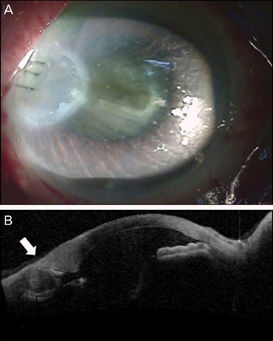
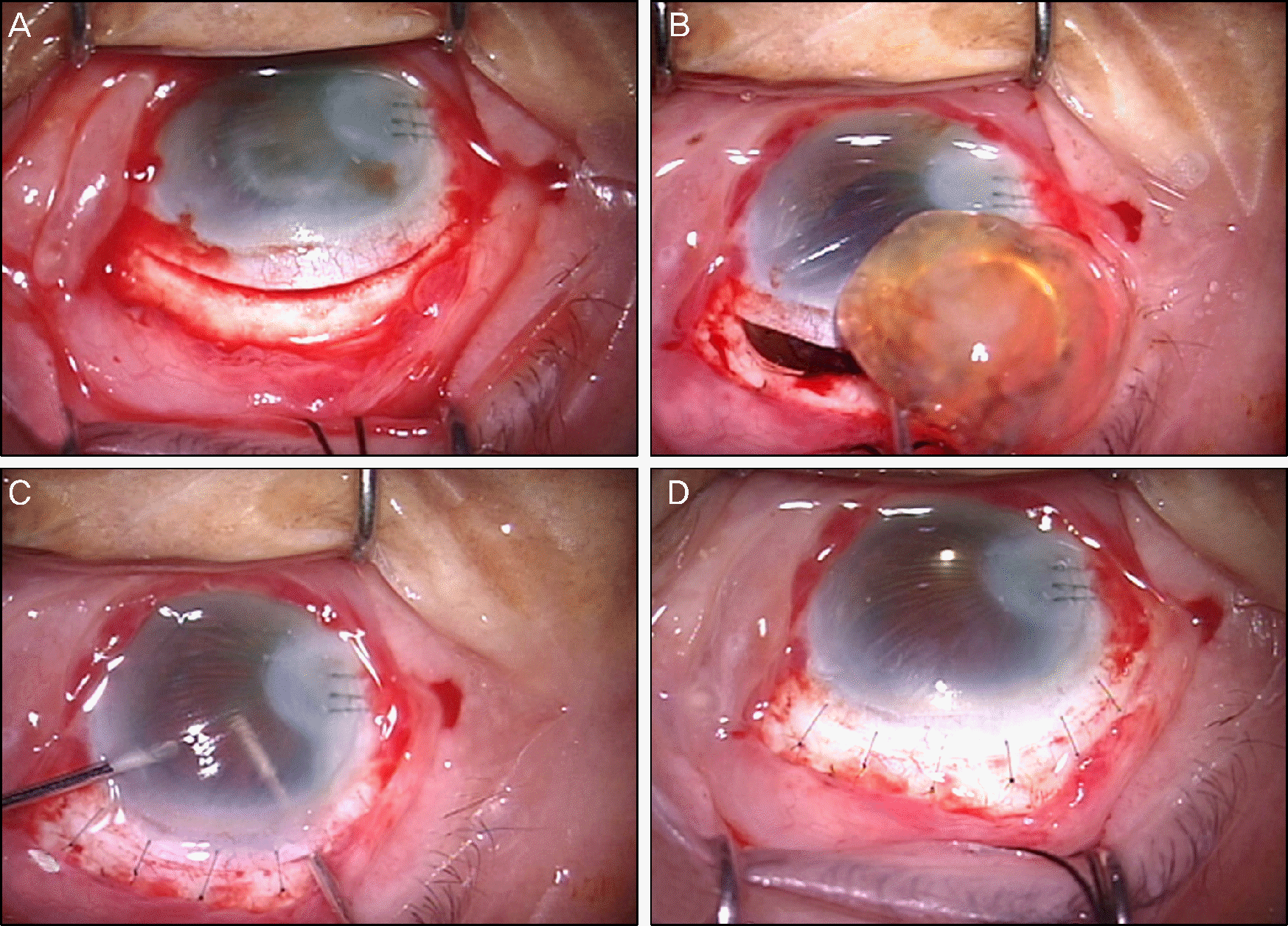
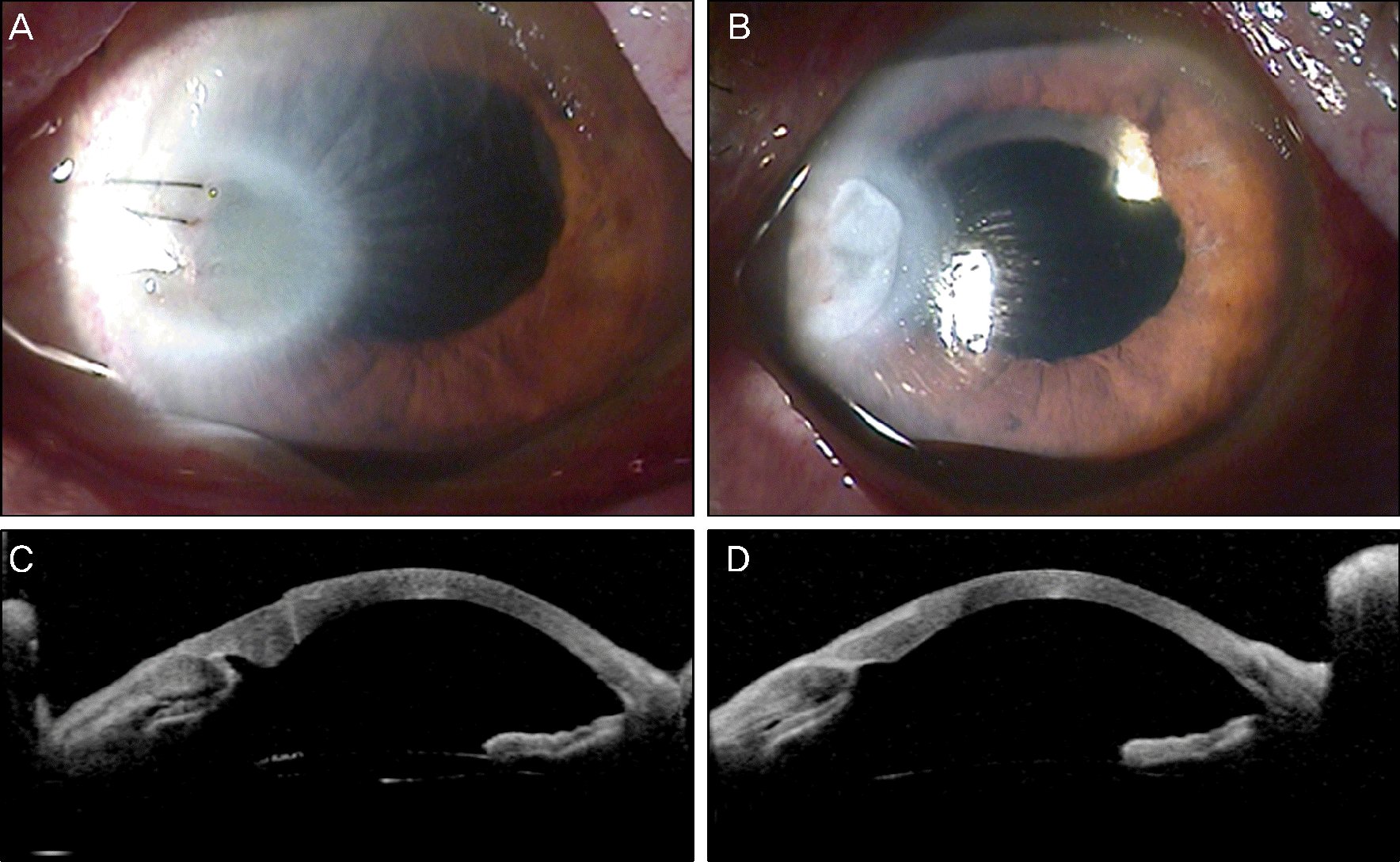
An 89-year-old female had phacoemulsification of a mature cataract in her right eye, and was transferred to our outpatient clinic after development of a thermal burn at the corneal incision site. On initial examination, visual acuity was light perception and slit-lamp examination revealed diffuse, severe corneal edema, and a 3.0 × 3.0 mm-sized epithelial defect with severe stromal opacity around the incision site. Extracapsular cataract extraction through superior scleral incision was performed with posterior chamber implantation of a 3-piece hydrophobic acrylic intraocular lens (IOL). Topical steroids as well as hypertonic saline were used to manage corneal edema postoperatively. One month post-operatively, her best corrected visual acuity was 0.06 and slit-lamp examination showed markedly decreased corneal edema and epithelial defect. Three months postoperatively, her best corrected visual acuity was 0.2, the IOL was centered in the capsular bag, and corneal edema nearly disappeared with remnant moderate corneal opacities.
References
1. Paik HJ, Song HJ, Shyn KH. 2007 Survey for KSCRS Members - current trends in cataract surgery in Korea. J Korean Ophthalmol Soc. 2009; 50:1624–31.
2. Brinton JP, Adams W, Kumar R, Olson RJ. Comparison of thermal features associated with 2 phacoemulsification machines. J Cataract Refract Surg. 2006; 32:288–93.

3. Bradley MJ, Olson RJ. A survey about phacoemulsification incision thermal contraction incidence and causal relationships. Am J Ophthalmol. 2006; 141:222–4.

4. Ernest P, Rhem M, McDermott M, et al. Phacoemulsification con-ditions resulting in thermal wound injury. J Cataract Refract Surg. 2001; 27:1829–39.

5. Mencucci R, Ambrosini S, Ponchietti C, et al. Ultrasound thermal damage to rabbit corneas after simulated phacoemulsification. J Cataract Refract Surg. 2005; 31:2180–6.

6. Davis PL. Phaco transducers: basic principles and corneal thermal injury. Eur J Implant Refract Surg. 1993; 5:109–12.

7. Sugar A, Schertzer RM. Clinical course of phacoemulsification wound burns. J Cataract Refract Surg. 1999; 25:688–92.

8. Kelman CD. Phaco-emulsification and aspiration. A new technique of cataract removal. A preliminary report. Am J Ophthalmol. 1967; 64:23–35.
9. Jun B, Berdahl JP, Kim T. Thermal study of longitudinal and torsional ultrasound phacoemulsification: tracking the temperature of the corneal surface, incision, and handpiece. J Cataract Refract Surg. 2010; 36:832–7.
10. Osher RH, Injev VP. Thermal study of bare tips with various system parameters and incision sizes. J Cataract Refract Surg. 2006; 32:867–72.

11. Allain JC, Le Lous M, Cohen-Solal , et al. Isometric tensions developed during the hydrothermal swelling of rat skin. Connect Tissue Res. 1980; 7:127–33.

12. Dada T, Sharma N, Vajpayee RB, Dada VK. Conversion from phacoemulsification to extracapsular cataract extraction: incidence, risk factors, and visual outcome. J Cataract Refract Surg. 1998; 24:1521–4.

13. Mercieca K, Brahma AK, Patton N, McKee HD. Intraoperative conversion from phacoemulsification to manual extracapsular cataract extraction. J Cataract Refract Surg. 2011; 37:787–8.

14. Bourne RR, Minassian DC, Dart JK, et al. Effect of cataract surgery on the corneal endothelium: modern phacoemulsification compared with extracapsular cataract surgery. Ophthalmology. 2004; 111:679–85.
15. Kim MS, Woo HM. Efficacy and safety of commercial used vis-coelastics: Healon®, Provisc®, Viscorneal®, Hyal 2000®, Biolon®, Viscoat®. J Korean Ophthalmol Soc. 2000; 41:1726–33.
16. Koch DD, Liu JF, Glasser DB, et al. A comparison of corneal endothelial changes after use of Healon or Viscoat during phacoemulsification. Am J Ophthalmol. 1993; 115:188–201.

17. Huang Y, Meek KM, Ho MW, Paterson CA. Anaylsis of bi-refringence during wound healing and remodeling following alkali burns in rabbit cornea. Exp Eye Res. 2001; 73:521–32.

18. Waterbury L, Kunysz EA, Beuerman R. Effects of steroidal and non-steroidal anti-inflammatory agents on corneal wound healing. J Ocul Pharmacol. 1987; 3:43–54.

19. Stahl JH, Miller DB, Conway BP, Campochiaro PA. Dexamethasone and indomethacin attenuate cryopexy. Induced breakdown of the blood-retinal barrier. Graefes Arch Clin Exp Ophthalmol. 1987; 225:418–20.
20. Sugar A, Bokosky JE, Meyer RF. A randomized trial of topical corticosteroids in epithelial healing after keratoplasty. Cornea. 1984-1985; 3:268–71.

21. Rask R, Jensen PK, Ehlers N. Healing velocity of corneal epithelium evaluated by computer. The effect of topical steroid. Acta Ophthalmol Scand. 1995; 73:162–5.
22. Lu Y, Fukuda K, Liu Y, et al. Dexamethasone inhibition of IL-1-in-duced collagen degradation by corneal fibroblasts in three-dimensional culture. Invest Ophthalmol Vis Sci. 2004; 45:2998–3004.

23. Kondo Y, Fukuda K, Adachi T, Nishida T. Inhibition by a selective IkB kinase-2 inhibitor of interleukin-1-induced collagen degradation by corneal fibroblasts in three-dimensional culture. Invest Ophthalmol Vis Sci. 2008; 49:4850–7.
24. Kupferman A, Leibowitz HM. Anti-inflammatory effectiveness of topically administered corticosteroids in the cornea without epithelium. Invest Ophthalmol. 1975; 14:252–5.
Figure 1.
Slit lamp photograph (A) and anterior segment optical coherence tomography (B) of an 89-year-old woman with thermal burn of the temporal cornea (white arrow).


 PDF
PDF ePub
ePub Citation
 Print
Print


 XML Download