

 PDF
PDF ePub
ePub Citation
 Print
Print


Abstract
Purpose
To report the results obtained after fixation of the eyeball to the periosteum over the posterior lacrimal crest in 5 cases of inveterate exotropia.
Methods
From September 2011 to January 2013, 5 patients with inveterate exotropia and a history of surgery for exotropia underwent fixation of the eyeball to the periosteum over the posterior lacrimal crest.
Results
The mean preoperative exotropia of 35 ± 10.61 PD changed to esotropia of 5.8 ± 17.28 PD at the one week post-operative visit and exotropia of 13.2 ± 11.34 PD at the final postoperative visit (mean, 6.05 months after surgery). At the final postoperative visit, 2 patients who had ocular adhesion due to trauma showed 25 PD exotropia. None of the patients showed injuries to the lacrimal system, but 2 patients showed conjunctival granuloma.
References
1. Von Noorden GK. Binocular vision and ocular motility. 3rd ed.St. Louis: CV Mosby;1985. p. 369.
2. Cho YA, Rah SH, Kim MM, Lee JY. Vertical rectus muscles transposition in large exotropia with medial rectus muscle transection following endoscopic sinus surgery. Korean J Ophthalmol. 2008; 22:104–10.

3. Thacker NM, Velez FG, Demer JL, Rosenbaum AL. Strabismic complications following endoscopic sinus surgery: diagnosis and surgical management. J AAPOS. 2004; 8:488–94.
4. Rosenbaum AI, Santiago AP. Clinical strabismus management principles and surgical technique. Philadelphia: WB Saunders;1999. p. 252.
5. Helveston EM. Muscle transposition procedures. Surv Ophthalmol. 1972; 16:92–7.
6. Helveston EM. Atlas of Strabismus Surgery. 3rd ed.St. Louis: CV Mosby;1985. p. 254–9.
7. Von Noorden GK. Binocular vision and ocular motility. 5th ed.St. Louis: CV Mosby;1996. p. 422.
8. Peter LC. The use of the superior oblique as an internal rotator in third nerve paralysis. Trans Am Ophthalmol. 1933; 31:232–7.
9. Jackson E. Surgery of the eye. 3rd ed.Vol. 1. New York: Grune & Stratton;1952. p. 405.
10. Scott AB. Transposition of the superior oblique. Am Orthopt J. 1977; 27:11–4.
11. Morad Y, Kowal L, Scott AB. Lateral rectus muscle disinsertion and reattachment to the lateral orbital wall. Br J Ophthalmol. 2005; 89:983–5.
12. Saunders RA, Rogers GL. Superior oblique transposition for third nerve palsy. Ophthalmology. 1982; 89:310–6.
13. Salazar-León JA, Ramírez-Ortíz MA, Salas-Vargas M. The surgical correction of paralytic strabismus using fascia lata. J Pediatr Ophthalmol Strabismus. 1998; 35:27–32.
14. Srivastava KK, Sundaresh K, Vijayalakshmi P. A new surgical technique for ocular fixation in congenital third nerve palsy. J AAPOS. 2004; 8:371–7.
15. Sharma P, Gogoi M, Kedar S, Bhola R. Periosteal fixation in third-nerve palsy. J AAPOS. 2006; 10:324–7.
16. Goldberg RA, Rosenbaum AL, Tong JT. Use of apically based periosteal flaps as globe tethers in severe paralytic strabismus. Arch Ophthalmol. 2000; 118:431–7.
17. Moe KS. The precaruncular approach to the medial orbit. Arch Facial Plast Surg. 2003; 5:483–7.
18. Saxena R, Sinha A, Sharma P, et al. Precaruncular approach for medial orbital wall periosteal anchoring of the globe in oculomotor nerve palsy. J AAPOS. 2009; 13:578–82.
19. Suk KW, Park JM, Kim SS, Lee SJ. Periosteal fixation in bilateral total third nerve palsy. J Korean Ophthalmol Soc. 2008; 49:1203–8.
20. Parks MM. Causes of the adhesive syndrome. Symposium on Strabismus. Transactions of the New Orleans Academy of Ophthalmology. St. Louis: CV Mosby;1978. p. 269–79.
21. Wright KW. The fat adherence syndrome and strabismus after retina surgery. Ophthalmology. 1986; 93:411–5.
22. Johnson LV. Adherence syndrome; pseudoparalysis of the lateral or superior rectus muscles. AMA Arch Ophthalmol. 1950; 44:870–8.
23. Dunlap EA. Plastic implants in muscle surgery: a study of the possible use of plastic materials in the management of extraocular motility restrictions. Trans Am Ophthalmol Soc. 1967; 65:393–470.
24. Robinson MR, Lee SS, Rubin BI, et al. Topical corticosteroid therapy for cicatricial conjunctivitis associated with chronic graft-ver-sus-host disease. Bone Marrow Transplant. 2004; 33:1031–5.
25. Cruz OA. Evaluation of mitomycin to limit postoperative adhesions in strabismus surgery. J Pediatr Ophthalmol Strabismus. 1996; 33:89–92.
Figure 1.
Schematic drawings of surgical procedures. (A) Conjunctival incision is made at the nasal quadrant of the eyeball and tenon dissection is done. Medial rectus muscle is isolated using the muscle hook. (B) 1% lidocaine mixed with epi-nephrine (1:100000) is injected and transcaruncular conjunctival incision is made. (C) Dissection is performed posteriorly and na-sally to reach the periosteum with Colorado needle monopolar electrocautery. (D) Periosteum over posterior lacrimal crest is sutured with 5-0 double armed non absorbable braided polyester (Ethibond®). (E, F) 18 gauge needles are passed through the subtenon space and tracted laterally. (G) Passed needles are se-curely anchored to the sclera at the insertion site of medial rectus muscle. (H) The sutures are tightened. (I, J) Medial peril-imbal sclera is anchored with 6-0 black silk. The sutures passed through medial part of upper eyelid and tided.
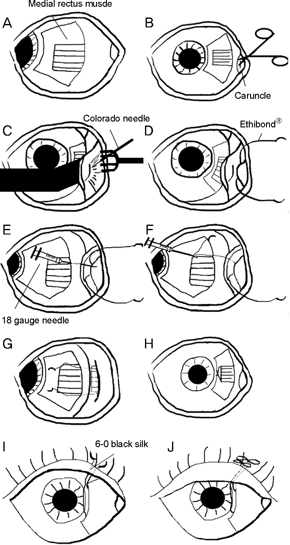
Figure 2.
Case 5. The preoperative 9 gaze photograph shows third nerve palsy in the left eye. The patient had ptosis and mild dilated pupil in the left eye and left exotropia in the primary position.
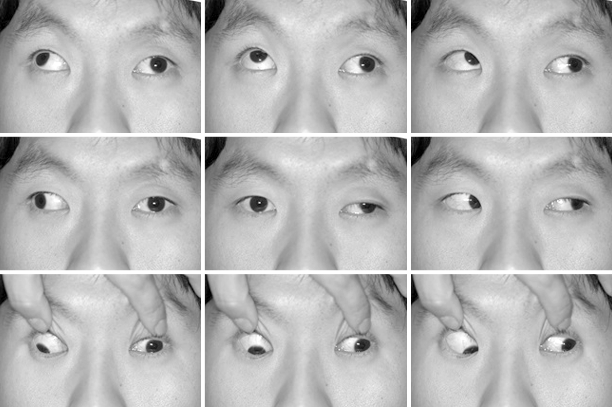
Figure 3.
Case 5. The 9 gaze photograph of postoperative 15 months shows mild exotropia in the primary position.
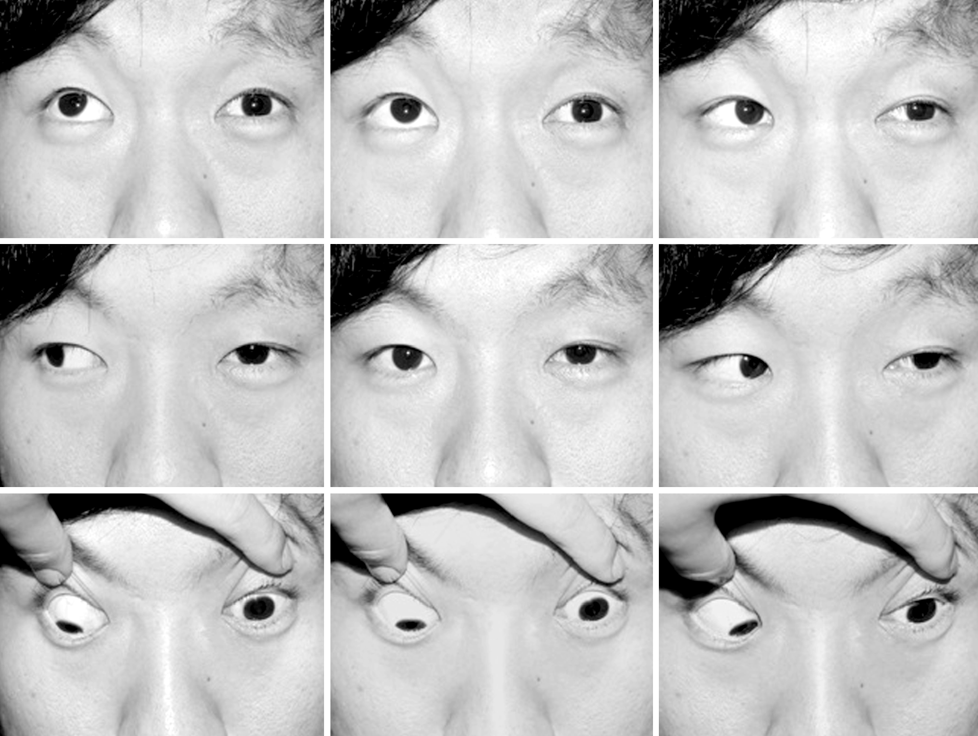
Table 1.
Characteristics of patients and results of surgery
 XML Download