

 PDF
PDF ePub
ePub Citation
 Print
Print


Abstract
Purpose
To research the advantage of using calcium plaque scissors in conjunctival flap surgery for calcified scleromalacia after cosmetic conjunctivectomy.
Methods
We analyzed 55 eyes that had undergone conjunctival flap surgery for calcified scleromalacia occurring after cosmetic conjunctivectomy. Surgical blade was used in 30 eyes (Group 1) and calcium plaque scissors in 25 eyes (Group 2). Time after conjunctivectomy, plaque size, operation time and visual acuity before and after the flap surgery were analyzed and compared. Additionally, necessity of additional scleral surgery was evaluated. Optical coherence tomography (OCT) of the sclera was performed both pre- and postoperatively and the results were compared.
Results
An average of 3.9 ± 1.0 years elapsed until conjunctival flap surgery and follow-up time was 5.2 ± 3.8 months. Post conjunctivectomy time and plaque size were similar in both groups (p = 0.87 and 0.49, respectively). The surgery time in Group 2 was shorter than in Group 1 (17.5 ± 6.3 minutes and 21.9 ± 8.5 minutes, respectively, p = 0.20). Uncorrected visual acuity was similar in both groups before and after conjunctival flap surgery (p = 0.53 and p = 0.20, respectively). In Group 1, one sclera transplantation and three OlogenTM insertion surgeries were performed as an additional scleral surgery. Based on OCT, calcium plaque scissors were confirmed as a new surgical tool for calcium plaque removal with minimal scleral damage.
References
1. Rhiu S, Shim J, Kim EK, et al. Complications of cosmetic wide conjunctivectomy combined with postsurgical mitomycin C application. Cornea. 2012; 31:245–52.

2. Leung TG, Dunn JP, Akpek EK, Thorne JE. Necrotizing scleritis as a complication of cosmetic eye whitening procedure. J Ophthalmic Inflamm Infect. 2013; 3:39.
3. Kwon HJ, Nam SM, Lee SY, et al. Conjunctival flap surgery for calcified scleromalacia after cosmetic conjunctivectomy. Cornea. 2013; 32:821–5.
4. Nguyen QD, Foster CS. Scleral patch graft in the management of necrotizing scleritis. Int Ophthalmol Clin. 1999; 39:109–31.
5. Hayasaka S, Noda S, Yamamoto Y, Setogawa T. Postoperative instillation of low-dose mitomycin C in the treatment of primary pterygium. Am J Ophthalmol. 1988; 106:715–8.
6. Chung DS, Cho BJ, Moon NJ. Mitomycin C single soaking during surgery for primary pterygium. J Korean Ophthalmol Soc. 1996; 37:927–33.
7. Shin HY, Kim MS, Chung SK. The development of scleromalacia after regional conjunctivectomy with the postoperative application of mitomycin C as an adjuvant therapy. Korean J Ophthalmol. 2013; 27:208–10.
8. Chen S, Noonan C. Scleral dellen complicating primary pterygium excision. Eye (Lond). 2000; 14:100–1.
9. Mitra S, Ganesh A, Shenoy R. Scleral dellen complicating primary pterygium excision. Eye (Lond). 2000; 14:924–5.
10. Moriarty AP, Crawford GJ, McAllister IL, Constable IJ. Severe corneoscleral infection. A complication of beta irradiation scleral necrosis following pterygium excision. Arch Ophthalmol. 1993; 111:947–51.
11. Solomon A, Kaiserman I, Raiskup FD, et al. Long-term effects of mitomycin C in pterygium surgery on scleral thickness and the conjunctival epithelium. Ophthalmology. 2004; 111:1522–7.
12. Dunn JP, Seamone CD, Ostler HB, et al. Development of scleral ulceration and calcification after pterygium excision and mitomycin therapy. Am J Ophthalmol. 1991; 112:343–4.
13. Hayasaka S, Iwasa Y, Nagaki Y, et al. Late complications after pterygium excision with high dose mitomycin C instillation. Br J Ophthalmol. 2000; 84:1081–2.
14. Rubinfeld RS, Pfister RR, Stein RM, et al. Serious complications of topical mitomycin-C after pterygium surgery. Ophthalmology. 1992; 99:1647–54.
15. Tsai YY, Lin JM, Shy JD. Acute scleral thinning after pterygium excision with intraoperative mitomycin C: a case report of scleral dellen after bare sclera technique and review of the literature. Cornea. 2002; 21:227–9.
16. Wan Norliza WM, Raihan IS, Azwa JA, Ibrahim M. Scleral melting 16 years after pterygium excision with topical Mitomycin C adjuvant therapy. Cont Lens Anterior Eye. 2006; 29:165–7.
17. Oh JH, Kim JC. Repair of scleromalacia with preserved scleral and amniotic membrane transplantation. J Korean Ophthalmol Soc. 2001; 42:810–6.
18. Sangwan VS, Jain V, Gupta P. Structural and functional outcome of scleral patch graft. Eye (Lond). 2007; 21:930–5.
19. Hsu WC, Ritch R, Krupin T, Chen HS. Tissue bioengineering for surgical bleb defects: an animal study. Graefes Arch Clin Exp Ophthalmol. 2008; 246:709–17.
20. Rosentreter A, Mellein AC, Konen WW, Dietlein TS. Capsule excision and Ologen implantation for revision after glaucoma drainage device surgery. Graefes Arch Clin Exp Ophthalmol. 2010; 248:1319–24.
Figure 1.
Intraocular lens scissors and calcium plaque scissors. (A) Shimowake Nucleus/intraocular lens (IOL) scissors. (B) Calcium plaque scissors, modified from Shimowake Nucleus/IOL scissors to have thinner tips.
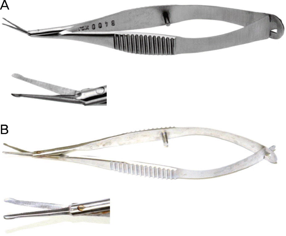
Figure 2.
The surgical procedure for calcified plaque removal and conjunctival flap surgery. (A) Firstly, the margin of calcified lesions are dissected using a surgical blade. (B) W ith calcium plaque scissors, calcified lesions are dissected just beneath the plaque. (C) After dissection, calcified plaque was cut by calcium plaque scissors. (D) The flap bed is prepared by scraping the epithelialized but nonvascularized area. (E) The conjunctival flap is pulled on the nonvascularized sclera and the subconjunctival tissue is released to decrease the flap contraction. (F) The conjunctival flap is sutured with enough 8-0 Vicryl stitches to fix it onto the sclera.
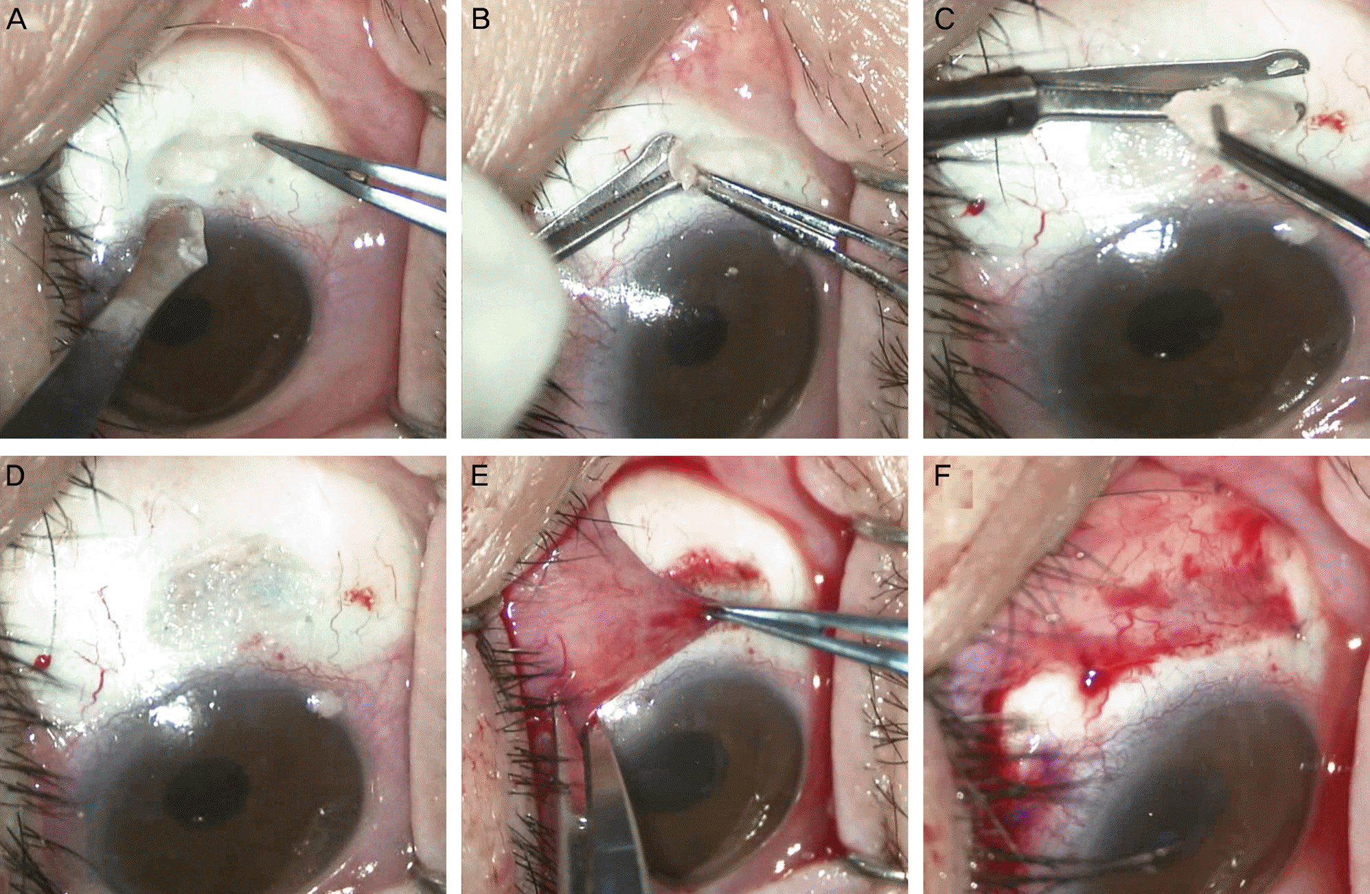
Figure 3.
The comparison of operation time between two groups. Box plot showing operation time in group 1 (knife) and group 2 (scissors). The mean operation time was 21.9 ± 8.5 minutes in group 1 (knife), and 17.5 ± 6.3 minutes in group 2 (scissors) (p = 0.03).
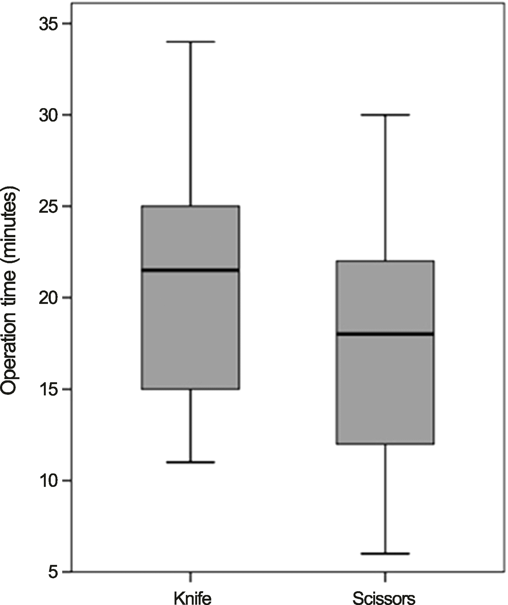
Figure 4.
Slit-lamp and fourier domain optical coherence tomography (FD-OCT) findings. (A, D, E) Preoperative slit-lamp and horizontal and vertical findings of FD-OCT images. Broad calcium plaque with dark margin is shown in FD-OCT (arrows). The image plane is indicated on photographs besides each OCT images. (B, F, G) One day after conjunctival flap surgery. Slit-lamp and horizontal and vertical FD-OCT images showed removed calcium plaque. (C) One month after conjunctival flap surgery.
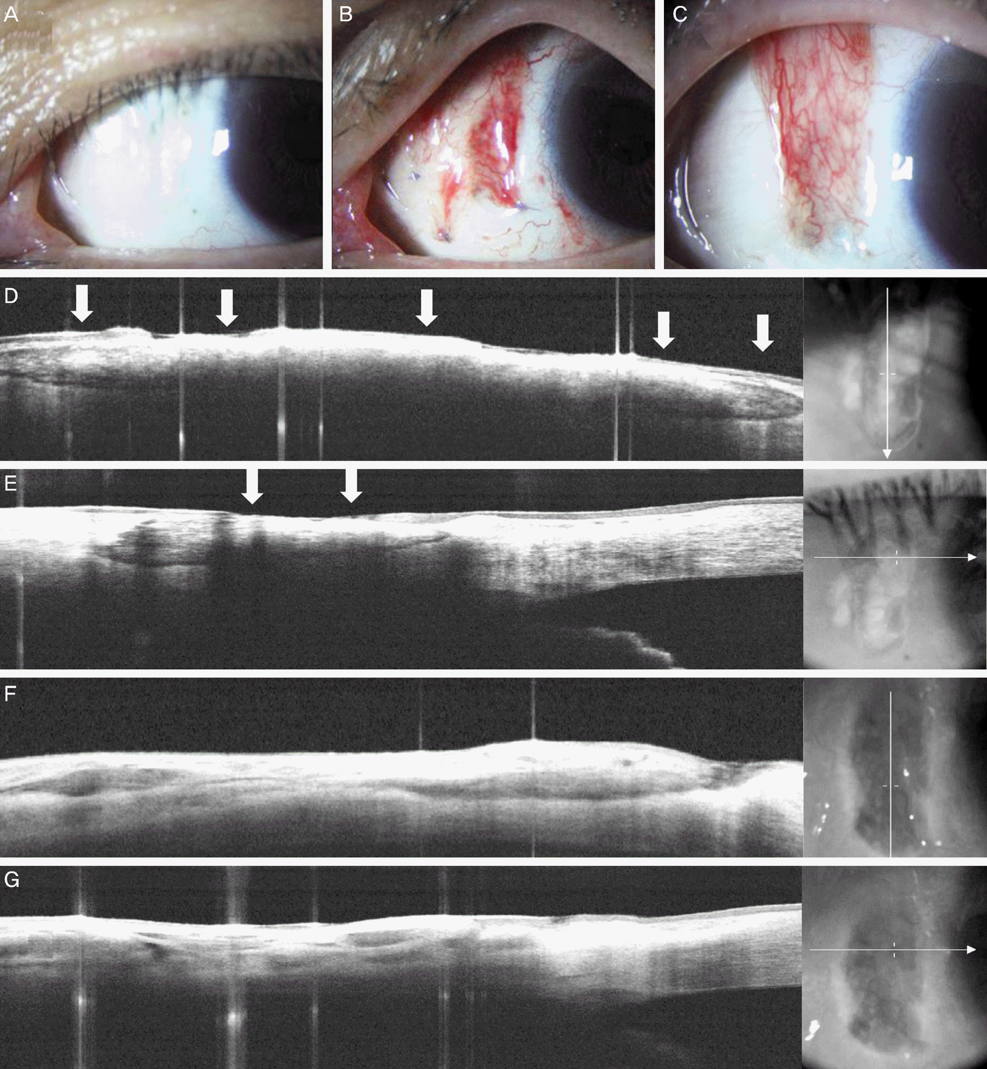
Table 1.
Baseline demographics
 XML Download