

 PDF
PDF ePub
ePub Citation
 Print
Print


Abstract
Purpose
To evaluate the association between ocular dominance, hand dominance and eye deviation in orthophoric and strabismus patients under general anesthesia during surgery.
Methods
The subjects were divided into 2 groups. Group 1 was composed of 38 patients who underwent strabismus surgery and group 2 was composed of 107 patients who underwent non-strabismus surgery under general anesthesia. Best corrected visual acuity (BCVA), dominant hand and fixating eye were obtained before surgery, and ocular dominance was assessed using the hole-in-the-card test. Under general anesthesia, we took a digital photo of both eyes, and the deviating eye was determined. Results: Under general anesthesia, the deviated eye showed no statistically significant correlation to the dominant eye and dominant hand in group I, respectively (p = 0.61, 0.74, respectively). In group II, there was no correlation between the deviated eye and the dominant eye (p = 0.65). The deviated eye also showed no correlation to the dominant hand in group II (p = 0.61). Conclusions: There was no correlation between the dominant and deviated eye under general anesthesia in the strabismus surgery group and the non-strabismus surgery group. Also, there was no correlation between the dominant hand and the deviated eye in patients under general anesthesia in the 2 groups.
References
1. BREININ GM. The position of rest during anesthesia and sleep; electromyographic observations. AMA Arch Ophthalmol. 1957; 57:323–6.

2. Duke-Elder S. System of ophthalmology. 6. St. Louis: C.V. Mosby Co.;1973. p. 96.
3. Coren S, Kaplan CP. Patterns of ocular dominance. Am J Optom Arch Am Acad Optom. 1973; 50:283–92.
4. Porac C, Coren S. Sighting dominance and binocular rivalry. Am J Optom Physiol Opt. 1978; 55:208–13.
5. Porac C, Coren S. Suppressive processes in binocular vision: ocular dominance and amblyopia. Am J Optom Physiol Opt. 1975; 52:651–7.
6. Worth C, Chavasse B. Squint. 9th ed.London: Bailliere, Tindal, and Cox;1959. p. 714.
7. Burford GE. Involuntary eyeball motion during anesthesia and sleep relationship to cortical rhythmic potentials. Anesth Anal. 1941; 20:191–9.
8. Roth A, Speeg-schatz CI. Eye muscle surgery: basic data operative techniques surgical stratergy. Swets and Zeitlinger publishers. Masson Paris. 1995; chap. 1, 4.
9. Omi E, Oguri K, Yoshiya I, Kitamura S. [Eye position of squinting eyes during general anesthesia (author's transl)]. Nihon Ganka Gakkai Zasshi. 1975; 79:540–6.
10. Romano P, Gabriel L. Intraoperative adjustment of eye muscle surgery. Correction based on eye position during general anesthesia. Arch Ophthalmol. 1985; 103:351–3.
11. Lee DS, Kim SY. Eye position of orthophoric patients under general anesthesia. J Korean Ophthalmol Soc. 2001; 42:1303–8.
12. Sidikaro Y, von Noorden GK. Observations in sensory heterotropia. J Pediatr Ophthalmol Strabismus. 1982; 19:12–9.
13. Havertape SA, Cruz OA, Chu FC. Sensory strabismus–eso or exo? J Pediatr Ophthalmol Strabismus. 2000; 38:327–30.
14. Min BM, Min WK, Lee KM, Kim YB. Clinical evaluation of sensory heterotropia. J Korean Ophthalmol Soc. 1989; 30:767–72.
15. Bielschowsky A. Über die relative Ruhelage der Augen. BerVers Deutsch Ophthal Ges. 1913; 39:67–79.
16. Chavasse FB. Worth's Squint or the Binorcular Reflexes and the Treatment of Strabismus. 7th ed.London: Bailliere, Tindall & Cox;1939. p. 519.
17. Knapp P. Divergent deviation. Allen JH, editor. Strabismus Ophthalmic Symposium II. 1st ed.St. Louis: Mosby;1958; chap. 17.
18. Fink WH. The dominant eye: its clinical significance. Arch Ophthalmol. 1938; 19:555–82.
19. Porac C, Coren S. The dominant eye. Psychol Bull. 1976; 83:880–97.
20. Mapp AP, Ono H, Barbeito R. What does the dominant eye dominate? A brief and somewhat contentious review. Percept Psychophys. 2003; 65:310–7.
21. Koo BS, Cho YA. The relationship of dominant eye, dominant hand, and deviated eye in strabismus. J Korean Ophthalmol Soc. 1996; 37:1277–82.
22. Cho KJ, Kim SY, Yang SW. The refractive errors of dominant and non-dominant eyes. J Korean Ophthalmol Soc. 2009; 50:275–9.
Figure 1.
Schematic illustration of eye position in the surgical plane of anesthesia. (A) Distance between temporal corneal limbus and corneal reflex in non-deviating eye. (B) Distance between nasal corneal limbus and corneal reflex in non-deviating eye. (C) Distance between nasal corneal limbus and corneal reflex in deviating eye. (D) Distance between temporal corneal limbus and corneal reflex in deviating eye.
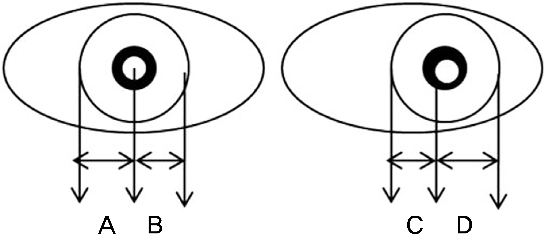
Table 1.
Characteristic of patients in strabismus and non-strabismus group at baseline
| Strabismus surgery (n = 38) |
Non-strabismus surgery (n = 107) |
p-value* |
||
|---|---|---|---|---|
| Ophthalmologic surgery (n = 61) | Non-ophthalmologic surgery (n = 46) | |||
| Age (years) | 7.71 ± 3.19 | 48.23 ± 23.72 | 33.54 ± 22.69 | 0.00* |
| Sex (n) | ||||
| Male | 23 | 29 | 21 | |
| Female | 15 | 32 | 25 | |
| Diagnosis | Exotropia 27 | Retinal disorder 44 | ||
| Esotropia 9 | Lens disorder 4 | |||
| Hypertropia 2 | Lid disorder 9 | |||
| Others 4 | ||||
| BCVA (decimal) | ||||
| Right | 0.92 ± 0.21 | 0.62 ± 0.37 | 0.98 ± 0.08 | 0.00* |
| Left | 0.90 ± 0.21 | 0.53 ± 0.37 | 0.98 ± 0.08 | 0.00* |
| Difference of BCVA (decimal) | 0.03 ± 0.12 | 0.10 ± 0.30 | 0.01 ± 0.03 | 0.01* |
Table 2.
Dominant eye & hand and deviated eye under general anesthesia in strabismus surgery group and non-strabismus surgery group
Table 3.
Association of deviated eye with dominant eye and dominant hand in strabismus surgery group and non-strabismus surgery group
| Strabismus surgery | Non-strabismus surgery | |||
|---|---|---|---|---|
| r ± SE | pvalue* | r ± SE | p-value* | |
| Dominant eye | 0.09 ± 0.16 | 0.74* | −0.44 ± 0.10 | 0.79* |
| Dominant hand | 0.06 ± 0.14 | 0.85* | 0.05 ± 0.13 | 0.54* |
 XML Download