

 PDF
PDF ePub
ePub Citation
 Print
Print


Abstract
Purpose
To report a case of intravitreal bevacizumab injection for choroidal neovascularization following direct laser photocoagulation for central serous chorioretinopathy.
Case summary
A 44-year-old male patient with an 8-month history of metamorphopsia in his left eye visited our clinic and was diagnosed with central serous chorioretinopathy after performing refraction, fundus examination, fluorescein angiography (FAG) and optical coherence tomography (OCT). After 1 month, laser photocoagulation of the leaking point observed on the FAG was performed. After 8 weeks following laser photocoagulation, visual acuity was reduced to 0.4, subretinal hemorrhage accompanied by choroidal neovascularization was observed on FAG and OCT, and an intravitreal bevacizumab injection was administered. After 4 weeks following the injection, macular edema and subretinal hemorrhage decreased, visual acuity increased to 1.0 and was maintained properly. However, after 2 years, the central serous chorioretinopathy recurred and after 3 months, healed spontaneously.
Conclusions
Intravitreal bevacizumab injection is a safe and effective treatment for secondary choroidal neovascularization occurring after direct laser photocoagulation for central serous chorioretinopathy. In addition, a single treatment can maintain the patient's status with no recurrence of choroidal neovascularization over a long-term period.
Figures and Tables
Figure 1
Initial fundus photographs and fluorescein angiographs. The right eye shows retinal pigment epithelium atrophy at the macula (A). The left eye shows elevated serous lesion involving the macula (B). FAG of the right eye shows atrophic tract and window defect around the macular area in the late phase (C). FAG of the left eye shows a smokestack appearance around the macular area in the late phase (D). FAG = fluorescein angiograph.
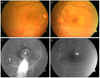
Figure 2
The baseline optical coherence tomography (OCT) of the left eye shows a neurosensory retinal detachment in the macular region (A), OCT of the left eye at four weeks after focal laser photocoagulation shows resolution of neurosensory retinal detachment (B).
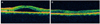
Figure 3
Fundus photograph of the left eye at two months after laser photocoagulation shows subretinal hemorrhage at the superotemporal area from the fovea involving macula (A). Fluorescein angiograph of the left eye at two months after laser photocoagulation shows a leakage with blockage by blood at the superotemporal area from the fovea (B). Optical coherence tomography of the left eye at two months after laser photocoagulation shows subretinal hemorrhage and choroidal neovascular membrane (C).
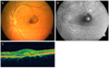
Figure 4
Fundus photograph of the left eye at five weeks after intravitreal bevasizumab injection shows decreased subretinal hemorrhage at the superotemporal area from the fovea (A). Fluorescein angiograph of the left eye at five weeks after intravitreal bevasizumab injection shows stained lesion without leakage at the superotemporal area from the fovea (B).
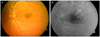
Figure 5
OCT of the left eye at two years after intravitreal bevasizumab injection shows a neurosensory retinal detachment in the macular region (A). OCT of the left eye at four months after a neurosensory retinal detachment shows resolution of neurosensory retinal detachment (B). OCT = optical coherence tomography.
References
1. Clais CM, Ober DM, Ciardella AP, Yannuzzi LA. Ryan SJ, editor. Central serous chorioretinopathy. Retina. 2006. Vol. 2:4th ed. Philadelphia: Elsevier-Mosby;1136–1161.
2. Burumcek E, Mudun A, Karacorlu S, Arslan MO. Laser photocoagulation for persistent central serous retinopathy: results of long-term follow-up. Ophthalmology. 1997. 104:616–622.
3. Gass JD. Photocoagulation treatment of idiopathic central serous choroidopathy. Trans Sect Ophthalmol Am Acad Ophthalmol Otolaryngol. 1977. 83(3 Pt 1):456–467.
4. Matsunaga H, Nangoh K, Uyama M, et al. [Occurrence of choroidal neovascularization following photocoagulation treatment for central serous retinopathy]. Nihon Ganka Gakkai Zasshi. 1995. 99:460–468.
5. Ha TW, Ham DI, Kang SW. Management of choroidal neovascularization following laser photocoagulation for central serous chorioretinopathy. Korean J Ophthalmol. 2002. 16:88–92.
6. Chan WM, Li KK, Liu DT, et al. Photodynamic therapy with verteporfin in laser-induced choroidal neovascularization. Am J Ophthalmol. 2003. 136:565–567.
7. Pikkel J, Rumelt S. Intravitreal bevacizumab for choroidal neovascularization secondary to laser photocoagulation for central serous chorioretinopathy. Eur J Ophthalmol. 2012. 22:488–491.
8. Cakir M, Cekiç O, Yilmaz OF. Photodynamic therapy for iatrogenic CNV due to laser photocoagulation in central serous chorioretinopathy. Ophthalmic Surg Lasers Imaging. 2009. 40:405–408.
9. Gilbert CM, Owens SL, Smith PD, Fine SL. Long-term follow-up of central serous chorioretinopathy. Br J Ophthalmol. 1984. 68:815–820.
10. Chung SE, Kang JH, Kang SW. Chronic central serous chorioretinopathy: photodynamic therapy. J Korean Ophthalmol Soc. 2007. 48:279–284.
11. Oh J, Kwon OW, Kim MH, et al. Photodynamic therapy for chronic central serous chorioretinopathy: multicenter study of 65 cases. J Korean Ophthalmol Soc. 2009. 50:390–398.
 XML Download