

 PDF
PDF ePub
ePub Citation
 Print
Print


Abstract
Purpose
We investigated the surgical anatomy of the deep lateral orbital wall via dissection of Korean cadavers and analysis of the orbit in normal adults using computed tomography.
Methods
Twelve cadavers were used to determine the exact anatomic index of the orbital lateral wall, and computed tomography images of 20 patients were used for surgical anatomic measurements during deep lateral orbital wall decompression. Additionally, the anatomic indexes measured in the cadavers and in the computed tomography study were compared and analyzed.
Results
In the cadaver study, the mean distance from the orbital rim to the end of the superior orbital fissure was 36.7 ±1.98 mm, to the rim of the frontosphendoial suture was 18.2 ± 1.92 mm, and from the end of the superior orbital fissure to the inferior orbital fissure was 17.1 ± 1.19 mm. In the computed tomography study, the mean value from the orbital rim to the end of the superior orbital fissure was 39.2± 2.46 mm, and from the rim to the frontosphenoidal suture was 17.8 ± 1.56 mm.
References
1. Ruttum MS. Effect of prior orbital decompression on outcome of strabismus surgery in patients with thyroid ophthalmopathy. J AAPOS. 2000; 4:102–5.

2. Lyons CJ, Rootman J. Orbital decompression for disfiguring exophthalmos in thyroid orbitopathy. Ophthalmology. 1994; 101:223–30.
3. Garrity JA, Fatourechi V, Bergstralh EJ, et al. Results of transantral orbital decompression in 428 patients with severe Graves' ophthalmopathy. Am J Ophthalmol. 1993; 116:533–47.
4. Shorr N, Neuhaus RW, Baylis HI. Ocular motility problems after orbital decompression for dysthyroid ophthalmopathy. Ophthalmology. 1982; 89:323–8.
5. Abràmoff MD, Kalmann R, de Graaf ME, et al. Rectus extraocular muscle paths and decompression surgery for Graves orbitopathy: mechanism of motility disturbances. Invest Ophthalmol Vis Sci. 2002; 43:300–7.
6. Goldberg RA, Perry JD, Hortaleza V, Tong JT. Strabismus after balanced medial plus lateral wall versus lateral wall only orbital decompression for dysthyroid orbitopathy. Ophthal Plast Reconstr Surg. 2000; 16:271–7.
7. Ben Simon GJ, Wang L, McCann JD, Goldberg RA. Primary-gaze diplopia in patients with thyroid-related orbitopathy undergoing deep lateral orbital decompression with intraconal fat debulking: a retrospective analysis of treatment outcome. Thyroid. 2004; 14:379–83.
8. Chang EL, Piva AP. Temporal fossa orbital decompression for treatment of disfiguring thyroid-related orbitopathy. Ophthalmology. 2008; 115:1613–9.
9. Baldeschi L, MacAndie K, Hintschich C, et al. The removal of the deep lateral wall in orbital decompression: its contribution to exophthalmos reduction and influence on consecutive diplopia. Am J Ophthalmol. 2005; 140:642–7.
10. Limawararut V, Valenzuela AA, Sullivan TJ, et al. Cerebrospinal fluid leaks in orbital and lacrimal surgery. Surv Ophthalmol. 2008; 53:274–84.
11. Whitehouse RW, Batterbury M, Jackson A, Noble JL. Prediction of enophthalmos by computed tomography after ‘blow out’ orbital fracture. Br J Ophthalmol. 1994; 78:618–20.
12. Charterside DG, Chan CH, Whitehouse RW, Noble JL. Orbital volume measurement of pure blowout fractures of the orbital floor. Br J Ophthalmol. 1993; 77:100–2.
13. De santo LW. Transantral orbital decompression. Gorman CA, Wailer RR, Dyer JA, editors. The eye and orbit in thyroid disease. New York: Raven Press;1984. p. 231–51.
14. Goldberg RA, Kim AJ, Kerivan KM. The lacrimal keyhole, orbital door jamb, and basin of the inferior orbital fissure. Three areas of deep bone in the lateral orbit. Arch Ophthalmol. 1998; 116:1618–24.
15. Stabile JR, Trokel SM. Increase in orbital volume obtained by decompression in dried skulls. Am J Ophthalmol. 1983; 95:327–31.
16. Beden U, Edizer M, Elmali M, et al. Surgical anatomy of the deep lateral orbital wall. Eur J Ophthalmol. 2007; 17:281–6.
17. Kakizaki H, Nakano T, Asamoto K, Iwaki M. Posterior border of the deep lateral orbital wall–appearance, width, and distance from the orbital rim. Ophthal Plast Reconstr Surg. 2008; 24:262–5.
18. Kakizaki H, Takahashi Y, Asamoto K, et al. Anatomy of the superior border of the lateral orbital wall: surgical implications in deep lateral orbital wall decompression surgery. Ophthal Plast Reconstr Surg. 2011; 27:60–3.
19. Bakholdina VIu. Morphometric characteristics and typology of the human orbit. Morfologiia. 2008; 133:37–40.
Figure 1.
Right orbit. (A) Measuring the distance between the end of superior orbital fissure and the lateral orbit rim. (B) Measuring the distance between the end of inferior orbital fissure and lateral orbit rim.
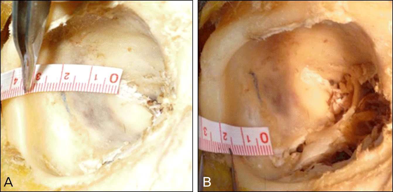
Figure 2.
The measurement of 5 surgical index of distance for deep lateral wall decompression in lateral orbit wall of cadavers. SOF = superior orbital fissure; FZS = frontozygomaticsuture; ZSS = zygomaticosphnoidal suture; GWS = greater wing of sphe-noid; IOF = inferior orbital fissure; ZFF = zygomaticofacial foramen; SOFD = superior orbital fissure distance; FSSD = frontosphenoidal suture distance; OFD = orbital fissure distance; IOFD = inferior orbital fissure distance; IFD = interfissure distance.
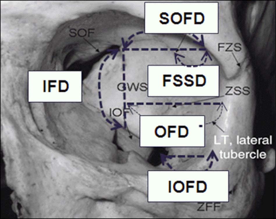
Figure 3.
The measurement of 4 surgical indexes for deep lateral wall decompression in orbital computed tomography. (A) The measurement of 3 surgical indexes in axial view of computed tomography. (B) The measurement of removal volume (arrow) of deep lateral wall decompression (GWSV) in axial view of computed tomography. GWST = greater wing of sphenoid thickness; GWSV = greater wing of sphenoid volume.
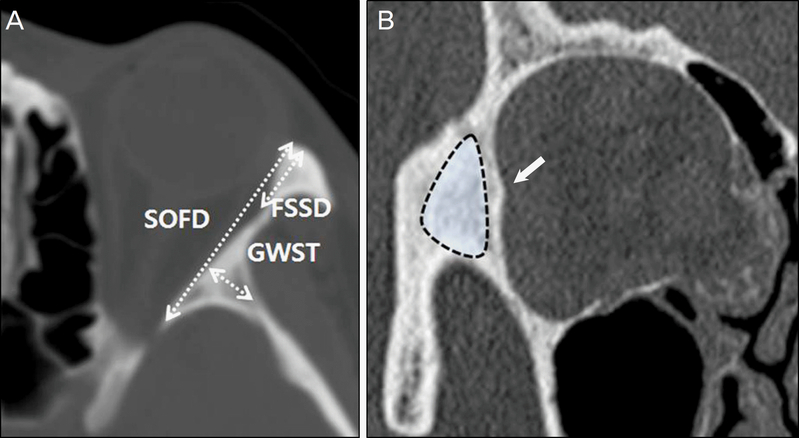
Table 1.
Sex and age of cadavers and computed tomography
| |
Cadavers |
Computed tomography |
||||
|---|---|---|---|---|---|---|
| Cases | Age (mean ± SD, yr) | p-value* | Cases | Age (mean ± SD, yr) | p-value* | |
| Male | 7 | 58.85 ± 12.52 | 0.38 | 10 | 55.82 ± 12.78 | 0.28 |
| Female | 5 | 65.58 ± 9.42 | 10 | 62.56 ± 8.77 | ||
Table 2.
Mean distance measured in lateral orbit wall of cadavers
| Index |
Distance (mean ± SD, mm) |
Significance p-value* | ||
|---|---|---|---|---|
| Male | Female | Total | ||
| SOFD | 37.89 ± 2.55 | 35.55 ± 1.58 | 36.72 ± 1.98 | 0.15 |
| FSSD | 18.78 ± 1.58 | 17.64 ± 2.25 | 18.21 ± 1.92 | 0.11 |
| IFD | 18.05 ± 0.98 | 16.25 ± 1.33 | 17.15 ± 1.19 | 0.09 |
| OFD | 37.25 ± 1.88 | 36.71 ± 2.18 | 36.98 ± 2.15 | 0.21 |
| IOFD | 16.12 ± 1.76 | 14.58 ± 1.95 | 15.35 ± 1.91 | 0.07 |
Table 3.
Mean value measured in lateral orbit wall from orbital computed tomography
| Index |
Mean value (mean ± SD) |
Significance p-value* | ||
|---|---|---|---|---|
| Male | Female | Total | ||
| SOFD (mm) | 39.99 ± 2.15 | 38.51 ± 2.47 | 39.25 ± 2.46 | 0.28 |
| FSSD (mm) | 18.11 ± 1.05 | 17.51 ±2.05 | 17.81 ± 1.56 | 0.25 |
| GWST (mm) | 5.54 ± 1.85 | 4.96 ± 1.35 | 5.25 ± 1.22 | 0.35 |
| GWSV (cm3) | 2.95 ± 1.28 | 2.73 ± 1.11 | 2.84 ± 0.94 | 0.21 |
 XML Download