

 PDF
PDF ePub
ePub Citation
 Print
Print


Abstract
Purpose
To describe a case of Wyburn-Mason Syndrome, which is characterized by arteriovenous malformations in the central nervous system and the retina.
Case summary:
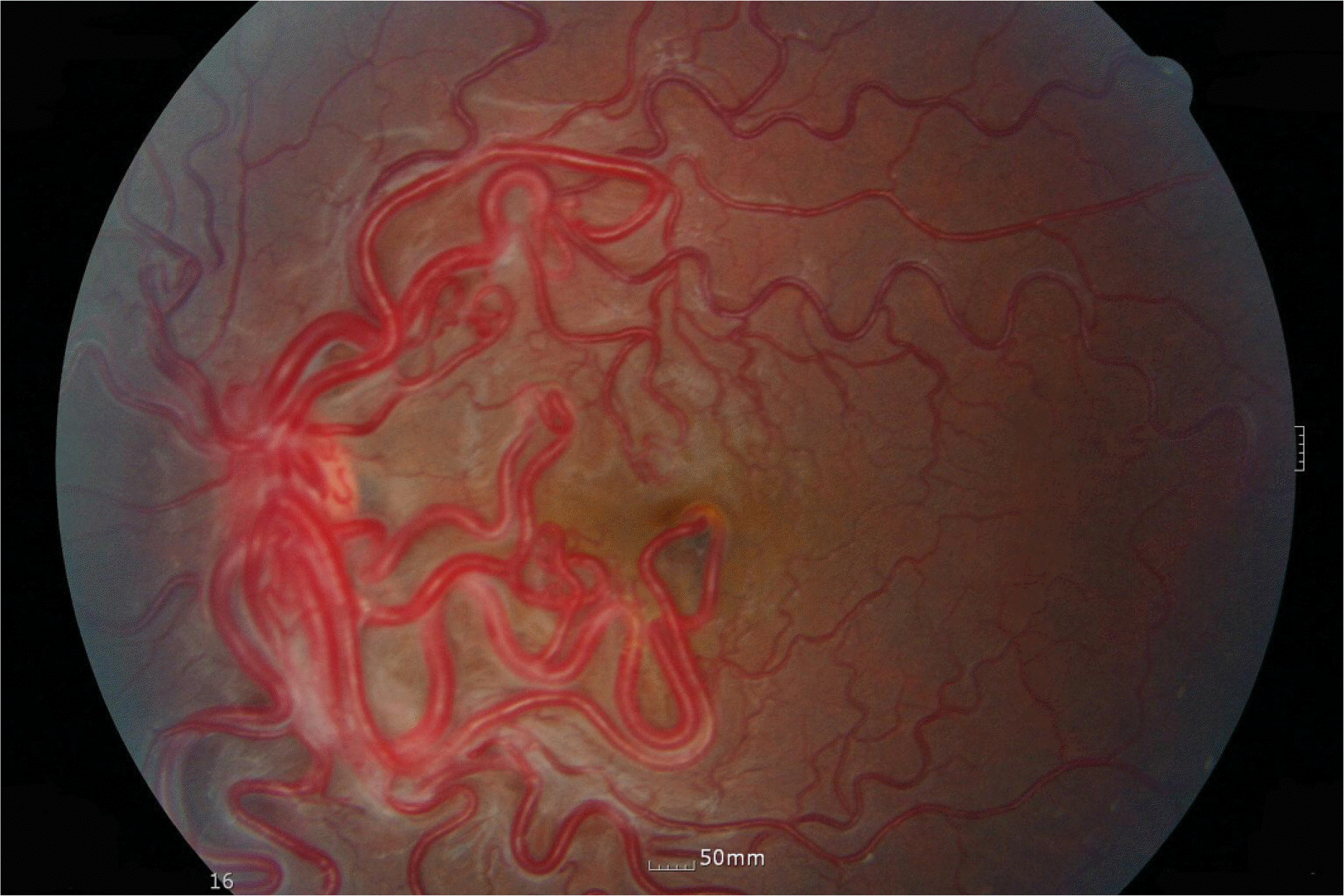
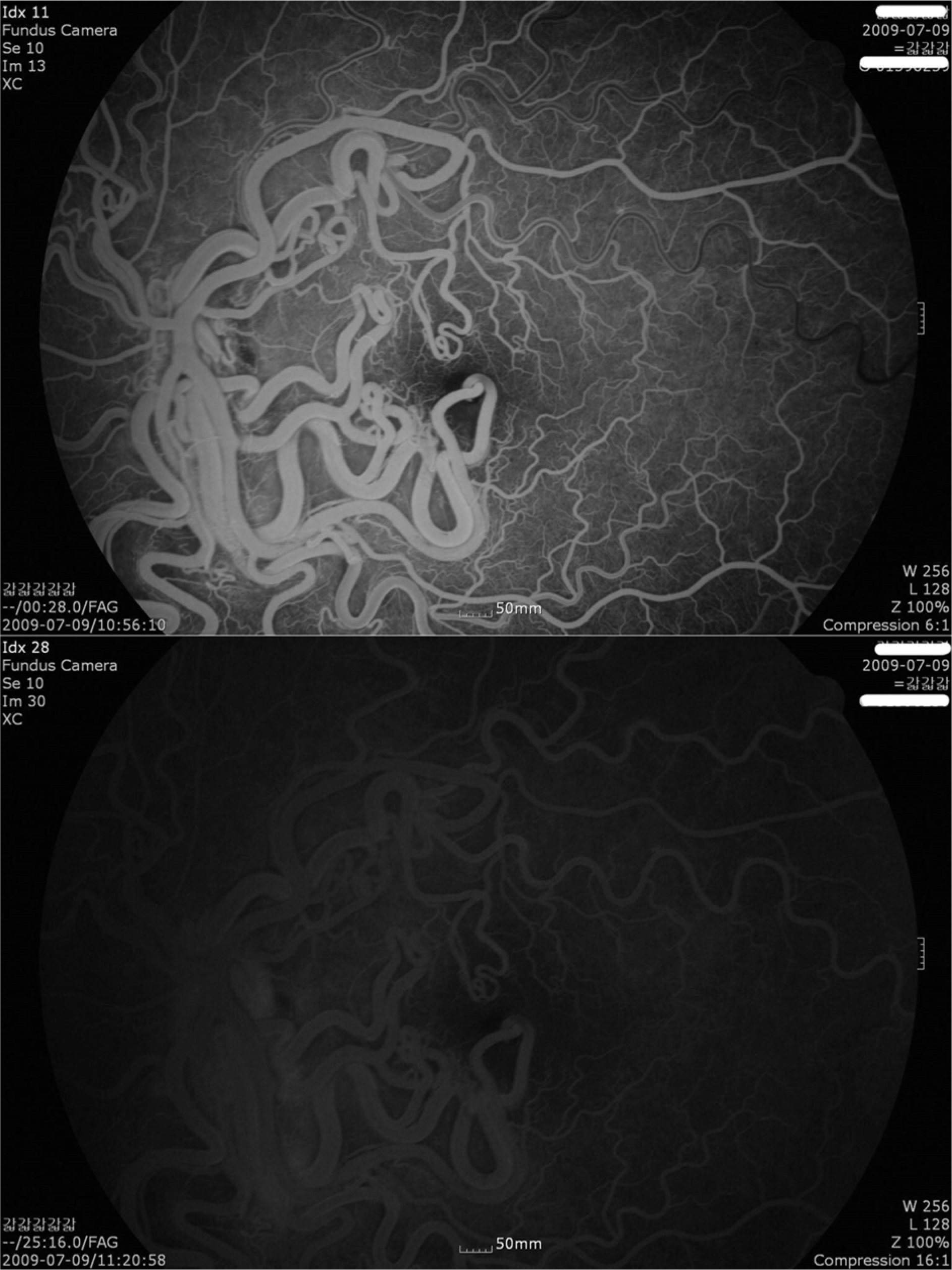
A 13-year-old girl who underwent neurosurgical intervention for intraventricular hemorrhage was referred to our ophthalmic clinic because brain angiogram and MRI finding revealed arteriovenous malformations (AVMs) that extended from the orbit and optic chiasm along the optic pathway. Fundus and fluorescein angiography of the left eye showed marked dilation and tortuosity of the retinal vessels. Fluorescein angiographic findings showed rapid filling of all vessels and no dye leakage.
References
1. Wyburn-Mason R. Arteriovenous aneurysm of midbrain and retina, facial naevi and mental changes. Brain. 1943; 66:163–203.
2. Bae HD, Kim BH, Shin DM, et al. A Case of Congenital aberrations Communication of the Retina. J Korean Ophthalmol Soc. 1981; 22:647–51.
3. Jang HS, Lee TK, Kim JW. A Case of Retinal Racemose aberrations. J Korean Ophthalmol Soc. 2001; 42:1232–5.
4. Bonnet P, Dechaume J, Blanc M. L'anevrisme cirsoide de la retine(Anevrysme racemeux). J de med de Lyon. 1937; 18:165–78.
5. Theron J, Newton TH, Hoyt WF. Unilateral retinocephalic vascular malformations. Neuroradiology. 1974; 7:185–96.

6. Bech K, Jensen OA. On the frequency of coexisting racemose aberrations of the retina and brain. Acta Psychiatr Neurol Scand. 1961; 36:47–56.
7. Goh D, Malik N N, Gilvarry A. Retinal racemose haemangioma directly communicating with a intramuscular facial cavernous haemangioma Br J Ophthalmol. 2004; 88:840–2.
8. Magnus H. Aneurysma arteriosovenosum retinale. Virchows Arch Pathol Anat Physiol. 1874; 60:38–43.
9. Carmeron ME, Greer CH. Congenital arteriovenous aneurysm of the retina, A post-mortem report. Brit Histol Ophthalmol. 1968; 52:768–72.
10. Noden DM. Cell movements and control of patterned tissue assembly during craniofacial development. J Craniofac Genet Dev Biol. 1991; 11:192–213.
11. Morgan MK, Johnston IH, De Silva M. Treatment of ophthalmofacial-hypothalamic arteriovenous malformation (Bonnet-Dechaume-Blanc syndrome). Case report. J Neurosurg. 1985; 63:794–6.
12. Reck SD, Zacks DN, Eibschitz-Tsimhoni M. Retinal and aberrations arteriovenous malformations: Wyburn-Mason syndrome. J Neuroophthalmol. 2005; 25:205–8.
13. Lee AW, Chen CS, Gailloud P, et al. Wyburn-Mason Syndrome Associated with Thyroid Arteriovenous Malformation: A First Case Report. Am J Neuroradiol. 2007; 28:1153–4.
Figure 1.
Angiograms of the left internal carotid artery (A: Anteroposterior view, B: Right antero-oblique view, C & D: Lateral view). There are AVMs that extends from the orbit and optic chiasm along the optic pathway to the midbrain (o: Ophthalmic artery, i: Internal carotid artery, AVM: AV malformations)

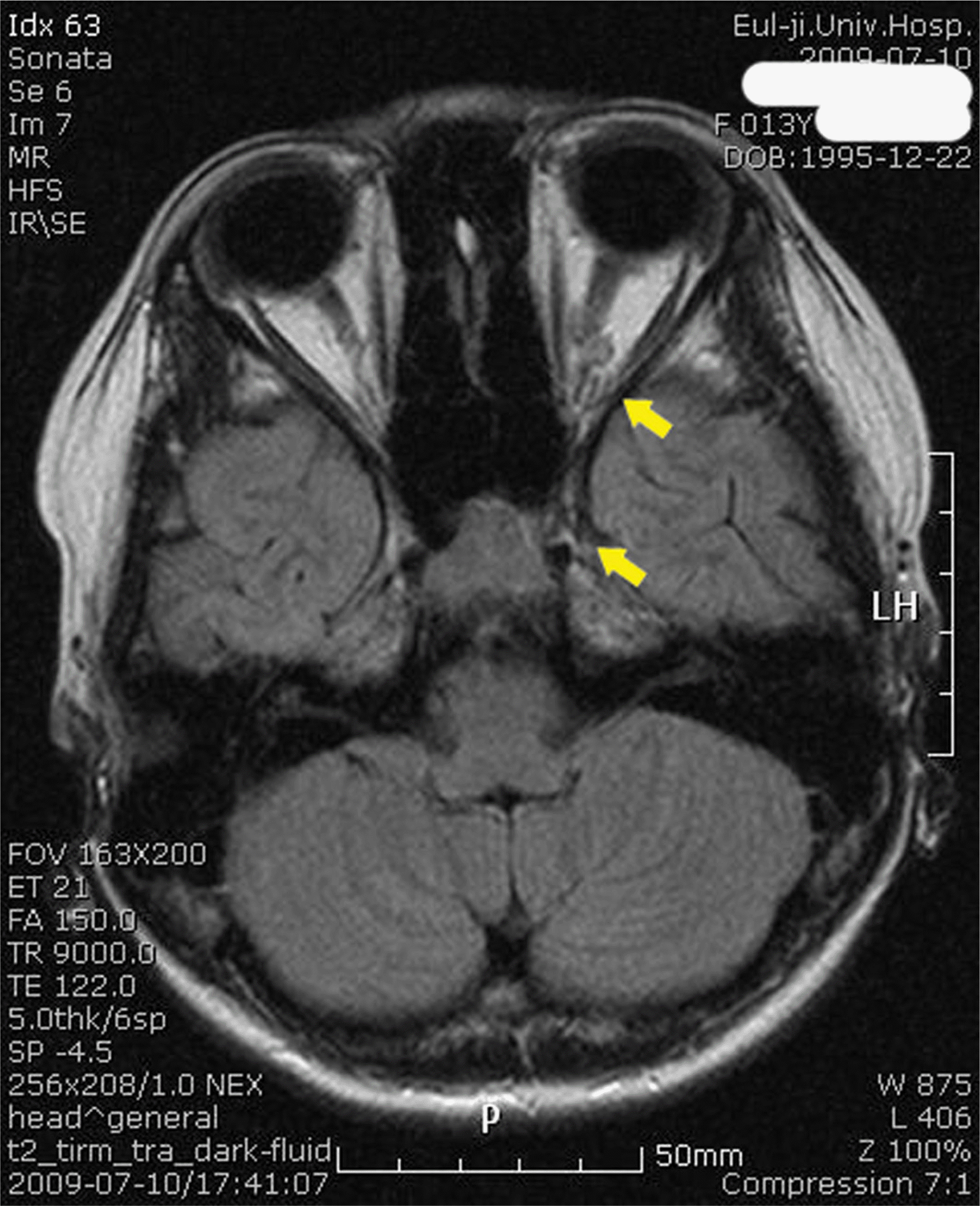
Figure 2.
Magnetic resonance imaging shows aggregated enhancing structures (yellow arrow) in the left central orbital space and suprasellar cistern, and around the supraclinoid internal carotid artery.
 XML Download