Abstract
Methods
A 52-year-old woman, who transferred from a private ophthalmic clinic to our hospital after a cataract operation due to a mass behind the iris, was evaluated. The patient had a dark brown mass with a smooth surface confirmed by ultrasonogram, CT, and MRI. The mass was removed by en bloc resection.
References
2. LoRusso FJ, Boniuk M. Font RL. Melanocytoma (magnocellular nevus) of the ciliary body. report of 10 cases and review of the literature. Ophthalmology. 2000; 107:795–800.
3. Al-Hinai A, Edelstein C, Burnier MN Jr. Unusual case of melanocytoma. Can J Ophthalmol. 2004; 39:461–3.

4. Capeans C, Pineiro A, Blanco MJ, et al. Ultrasound biomicroscopic findings in a cavitary melanocytoma of the ciliary body. Can J Ophthalmol. 2003; 38:501–3.
6. El-Harazi SM, Kellaway J, Font RL. Melanocytoma of the ciliary body diagnosed by fine-needle aspiration biopsy. Diagn Cytopathol. 2000; 22:394–7.

7. Mohamed MD, Gupta M, Parsons A, Rennie IG. Ultrasound biomicroscopy in the management of melanocytoma of the ciliary body with extrascleral extension. Br J Ophthalmol. 2005; 89:14–6.

8. Lee NK. A Case of melanocytoma of the optic disc. J Korean Ophthalmol Soc. 1975; 16:248–50.
9. Yoo JH, Joo MJ, Kim JS, Kwak HW. Vitreous seeding associated with melanocytoma of the optic disc. J Korean Ophthalmol Soc. 1996; 37:1761–4.
10. Lee J-Y, Kim SH. Non-invasive Diagnosis of Melanocytoma and 1 Case Report. J Korean Ophthalmol Soc. 2003; 44:517–22.
11. Shammas HJ, Minckler DS, Hulquist R, Sherins RS. Melanocytoma of the ciliary body. Ann Ophthalmol. 1981; 13:1381–3.
12. Fineman M, Eagle RC Jr, Shields JA, et al. Melanocytomalytic glaucoma in eyes with necrotic iris melanocytoma. Ophthalmology. 1998; 105:492–6.

13. Shields JA, Eagle RC Jr, Shields CL, Nelson LB. Progressive growth of an iris melanocytoma in a child. Am J Ophthalmol. 2002; 133:287–9.

14. Bhorade AM, Edward DP, Goldstein DA. Ciliary body melanocytoma with anterior segment pigment dispersion and elevated intraocular pressure. J Glaucoma. 1999; 8:129–33.

15. Shields CL, Naseripour M, Shields JA, et al. Custom-designed plaque radiotherapy for nonresectable iris melanoma in 38 patients: tumor control and ocular complications. Am J Ophthalmol. 2003; 135:648–56.

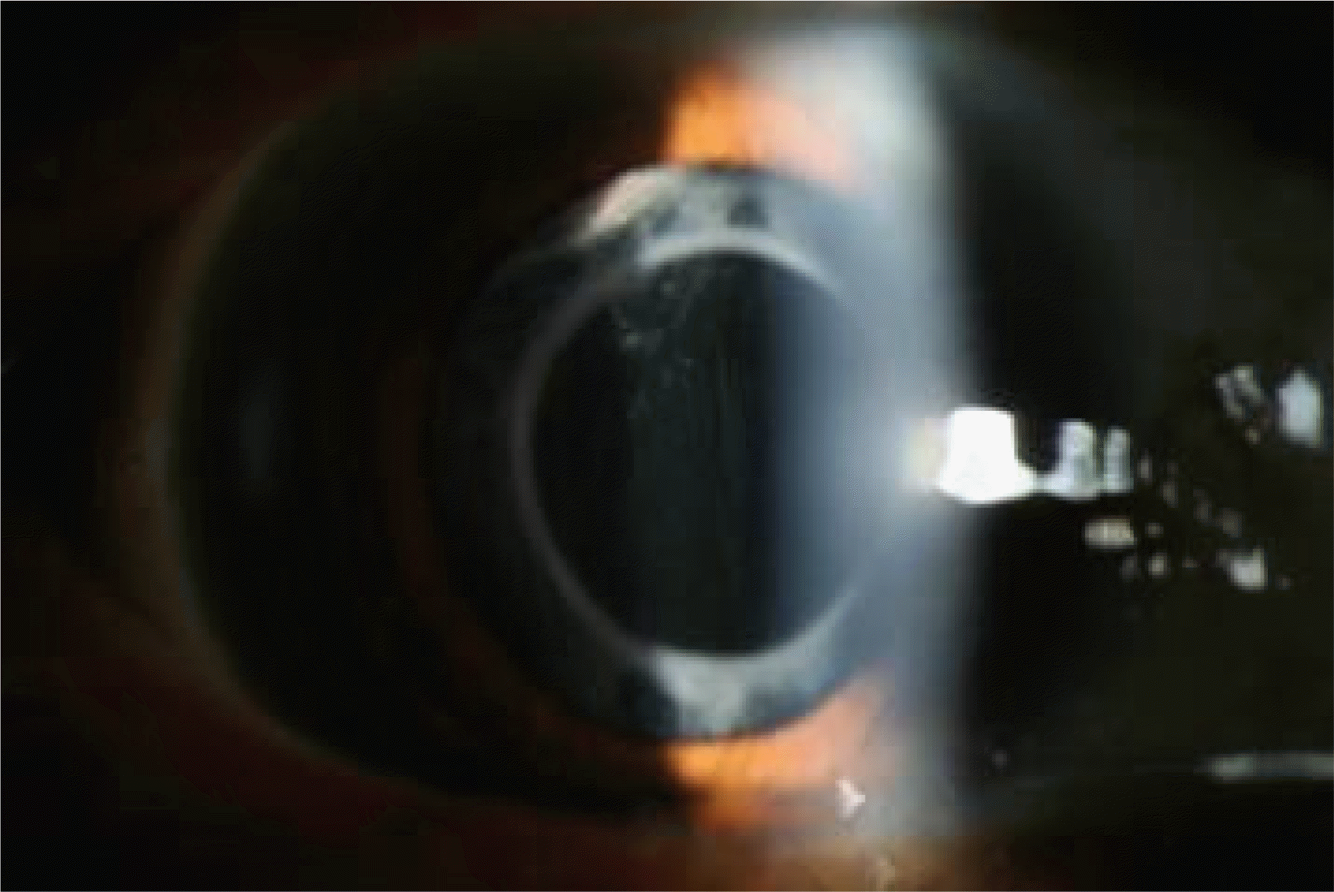
Figure 1.
Preoperative slit lamp examination of melanocytoma of the ciliary body showing at 11 o'clock position along dilated pupil margin.

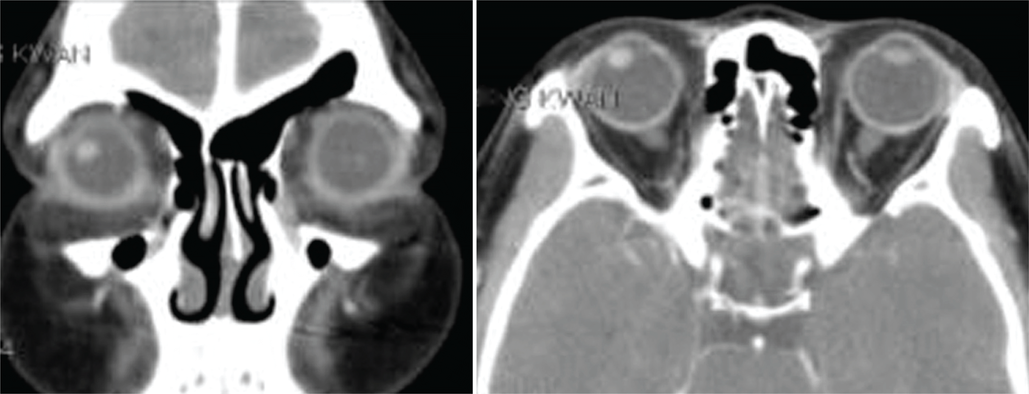
Figure 2.
Preoperative orbit CT finding: tumorous lesion at inferolateral aspect of the iris or ciliary body in the right eyeball.

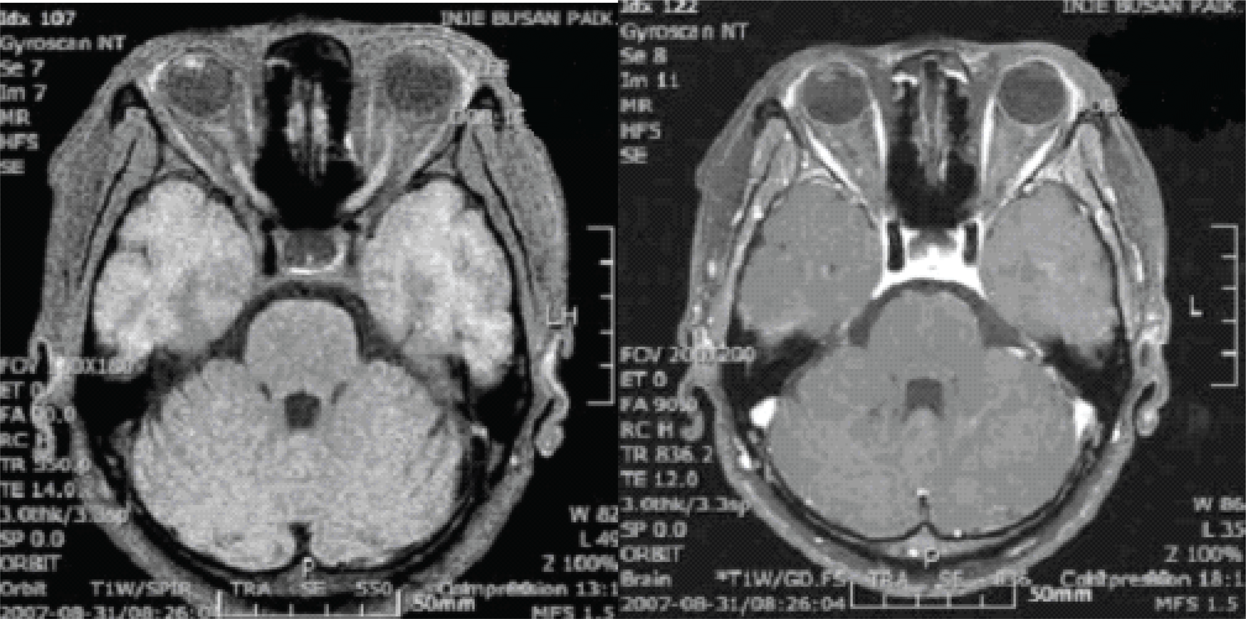
Figure 3.
Preoperative orbit MRI finding: well-defined, round mass with signal alteration and intense enhancement at the inferolateral aspect of the right iris or ciliary body.

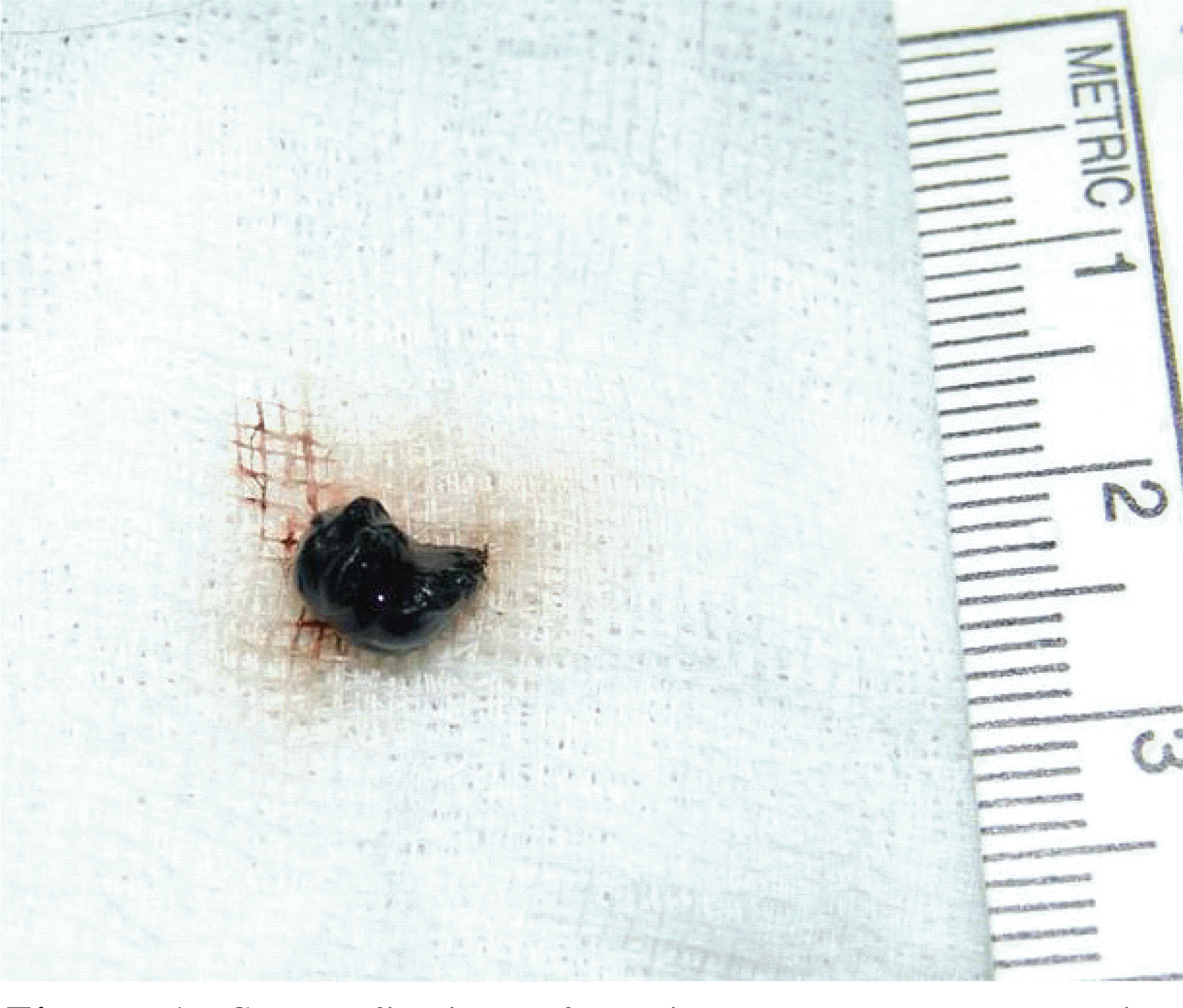
Figure 4.
Gross finding of excised tumor mass, which is dark brown, soft, and smooth surfaced and 0.5×0.5 ×0.5 cm in size.

Figure 5.
Microscopic appearance of melanocytoma of the ciliary body (A) (Histopathologic examination, ×40) tumor cells are polygonal melanocytes with small vesicular nuclei and abundant cytoplasm totally packed with melanin granules (B) tumor cells after bleaching preparation have abundant melanin pigment in the cytoplasm.


 PDF
PDF ePub
ePub Citation
 Print
Print


 XML Download