

 PDF
PDF ePub
ePub Citation
 Print
Print


Abstract
Purpose
To compare ocular higher order aberrations measured by two different aberrometers in a sample of normal eyes.
Methods
We included 41 normal eyes of Koreans in this study. Ocular aberration data were obtained through three measurements per eye using Zywave and OPD-Scan devices. Spherical equivalent and higher order aberrations calculated in the central 6 mm zone and expressed as root mean square (RMS) values were analyzed.
Results
A comparison of measurements between the Zywave and OPD-Scan devices demonstrated no statistically significant differences in the RMS values of total higher order aberration (p=0.11), but significant differences were detected in the RMS values of total spherical aberration, total coma and total trefoil (p<0.01).
References
1. Born M. Principles of optics. New York: Pergamon Press;1975. p. 464–6.
2. Rozema JJ, Van Dyck DE, Tassignon MJ. Clinical comparison of 6 aberrometers. Part 1: Technical specifications. J Cataract Refract Surg. 2005; 31:1114–27.

3. Rozema JJ, Van Dyck DE, Tassignon MJ. Clinical comparison of 6 aberrometers. Part 2: statistical comparison in a test group. J Cataract Refract Surg. 2006; 32:33–44.
4. Liang J, Grimm B, Goelz S, Bille JF. Objective measurement of wave aberrations of the human eye with the use of a Hartmann-Shack wavefront sensor. J Opt Soc Am A Opt Image Sci Vis. 1994; 11:1949–57.
5. Thibos LN. Principles of Hartmann-Shack aberrometry. J Refract Surg. 2000; 16:563–5.
6. MacRae S, Fujieda M. Slit Skiascopic-guided ablation using the Nidek Laser. J Refract Surg. 2000; 16:576–80.
7. Molebny V, Pallikaris IG, Naoumidis LP, et al. Retina ray-tracing technique for eye refraction mapping. SPIE Proc. 1997; 2971:175–83.
8. Navarro R, Moreno-Barriuso E. Laser ray-tracing method for optical testing. Opt Lett. 1999; 24:951–3.
9. Mrochen M, Kaemmerer M, Mierdel P, et al. Principles of Tscherning aberrometry. J Refract Surg. 2000; 16:570–1.
10. Mirshahi A, Buhren J, Gerhardt D, Kohnen T. In vivo and in vitro repeatability of Hartmann-Shack aberrometry. J Cataract Refract Surg. 2003; 29:2295–301.
11. Hament WJ, Nabar VA, Nuijts RM. Repeatability and validity of Zywave aberrometer measurements. J Cataract Refract Surg. 2002; 28:2135–41.
12. Durrie DS, Stahl ED. Comparing wavefront devices. Krueger RR, Applegate RA, MacRae SM, editors. Wavefront Customized Visual Correction: The Quest for Super Vision II. Thorofare, NJ: SLACK Inc;2004. 1:chap. 21.
13. Jeong JH, Kim MJ, Tchah HW. Clinical Comparison of Laser Ray Tracing Aberrometer and Shack-Hartmann Aberrometer. J Korean Ophthalmol Soc. 2006; 47:1911–9.
14. Altman DG, Bland JM. Measurements in medicine: the analysis of method comparison studies. Statistician. 1983; 32:307.
15. Bland JM, Altman DG. Statistical methods for assessing agreement between two methods of clinical measurements. Lancet. 1986; 1:307–10.
16. Bailey MD, Mitchell GL, Dhaliwal DK, et al. Patient satisfaction and visual symptoms after laser in situ keratomileusis. Ophthalmology. 2003; 110:1371–8.
17. Marcos S. Aberrations and visual performance following standard laser vision correction. J Refract Surg. 2001; 17:596–601.
18. Burakgazi AZ, Tinio B, Bababyan A, et al. Higher order aberrations in normal eyes measured with three different aberrometers. J Refract Surg. 2006; 22:898–903.
19. Rodriguez P, Navarro R, Gonzalez L, Hernández JL. Accuracy and reproducibility of Zywave, Tracey and experimental aberrometers. J Refract Surg. 2004; 20:810–7.
20. Cerviño A, Hosking SL, Montés-Micó R. Comparison of higher order aberrations measured by NIDEK OPD-Scan dynamic skiascopy and Zeiss WASCA Hartmann-Shack aberrometers. J Refract Surg. 2008; 24:790–6.
21. Kim DS, Narváez J, Krassin J, Bahjri K. Comparison of the VISX wavescan and NIDEK OPD-scan aberrometers. J Refract Surg. 2009; 25:429–34.
22. Liang CL, Juo SH, Chang CJ. Comparison of higher-order wavefront aberrations with 3 aberrometers. J Cataract Refract Surg. 2005; 31:2153–6.
Figure 1.
Repeatability Coefficient (μm)* of OPD-Scan TM and Zywave TM. This graphs show the distribution of the differences between each total higher order aberration measurement and the mean of three consecutive measurements in each of the 41 eyes. (Repeatibility coefficient: OPD-ScanTM=0.11 μm and ZywaveTM=0.06 μm)* Repeatability Coefficient (μm)=95% confidence interval for repeated measurements; HOA=higher order aberration.
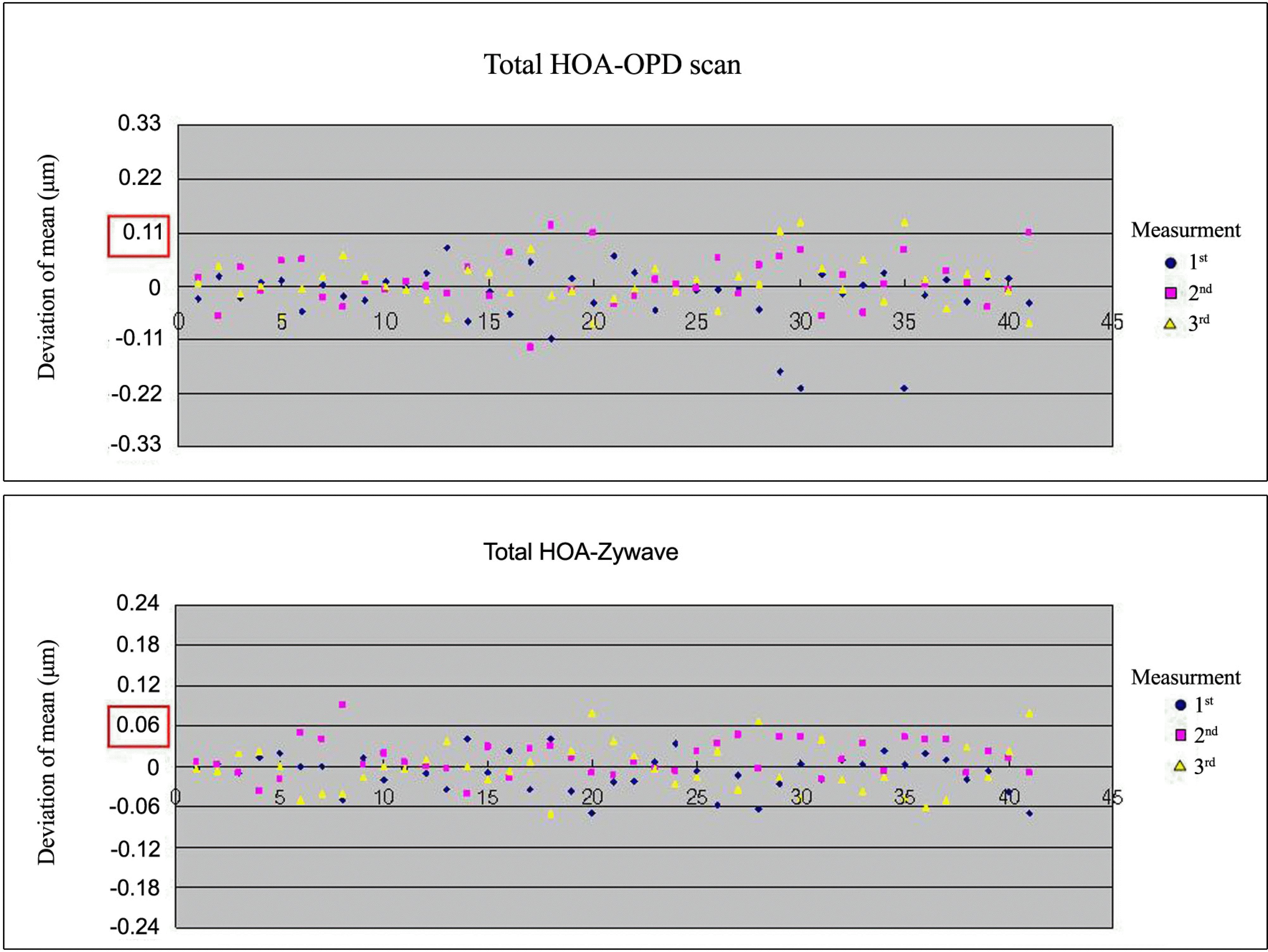
Figure 2.
Correlations of OPD-ScanTM and Zywave TM. HOA=higher order aberration; SA=spherical aberration; r=Pearson correlation coefficient (p<0.01).
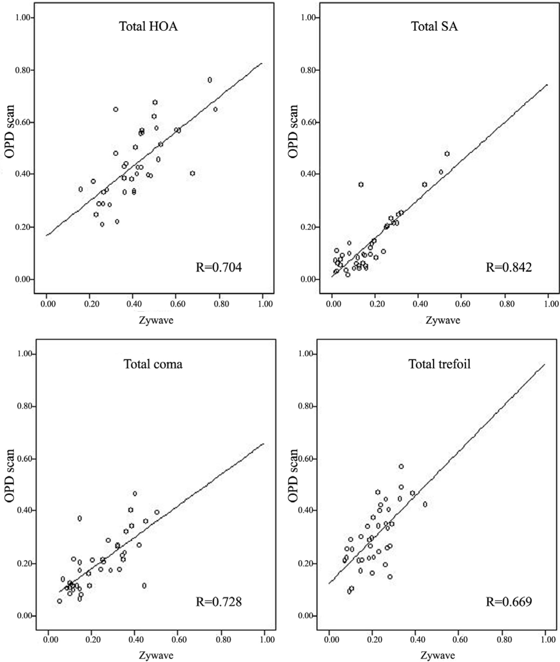
Table 1.
Comparisons of spherical equivalent of manifest refraction, OPD-Scan TM and Zywave TM
| MR* | OPD scan TM | Zywave TM | OPD scan TM-MR*(pair t-test) | Zywave TM-MR*(pair t-test) | OPDscan TM-Zywave TM (pair t-test) | |
|---|---|---|---|---|---|---|
| Spherical equivalent (Diopters) | −0.26±0.75 | −0.21±0.85 | +0.11±0.83 | 0.05±0.36(p=0.11) | 0.37±0.33(p<0.01) | −0.31±0.15(p<0.01) |
Table 2.
Repeatability Coefficient (μm)* of OPD-Scan TM and Zywave TM
| OPD scan TM | ZywaveTM | |
|---|---|---|
| Total higher order aberration (μm) | 0.11 | 0.06 |
| Total Spherical aberration (μm) | 0.12 | 0.05 |
| Total Coma (μm) | 0.13 | 0.06 |
| Total Trefoil (μm) | 0.13 | 0.08 |
Table 3.
Comparisons of higher order aberrations between OPD-Scan TM and Zywave TM
 XML Download