

 PDF
PDF ePub
ePub Citation
 Print
Print


Abstract
Purpose
To compare the retinal nerve fiber layer (RNFL) thickness among normal children, glaucoma suspects, and children with glaucoma, using optical coherence tomography (OCT).
Methods
Ninety‐ four eyes of 47 normal children, 62 eyes of 38 glaucoma suspects and 33 eyes of 21 children with glaucoma, from the ages of 5 to 15 years were examined at the Ophthalmology Center at the Inha University Hospital. The RNFL thickness was measured with OCT. Patient cooperation and signal strength of the OCT scans were assessed. The mean, superior, inferior, nasal, and temporal RNFL thicknesses were measured by OCT in all three groups.
Results
After adjustment by refractive error, the RNFL thicknesses of the mean, superior, inferior, nasal, and temporal areas were 110.8±10.1 µm, 140.1±17.5 µm, 134.1±14.8 µm, 79.9±14.5 µm, and 89.2±16.8 µm, respectively, in the normal group; 107.9±10.8 µm, 134.8±15.5 µm, 130.2±18.8 µm, 75.4±17.1 µm, and 91.2±18.7 µm, respectively, in the glaucoma suspect group; and 102.8±18.1 µm, 129.5±16.5 µm, 126.1±20.2 µm, 70.3±19.7 µm, and 85.1±16.9 µm, respectively, in the glaucoma patient group. There was a significant difference among the three groups in all locations except in the temporal area (p=0.003).
References
1. Mikelberg FS, Drance SM, Schulzer M. . The normal human optic nerve: Axon count and axon diameter distri-bution. Ophthalmology. 1989; 96:1325–8.
2. Sommer A, Miller NR, Pollack I. . The nerve fiber layer in the diagnosis of glaucoma. Arch Ophthalmol. 1977; 95:2149–56.

3. Quigley HA, Nickells RW, Kerrigan LA. . Retinal ganglion cell death in experimental glaucoma and after axotomy occurs by apoptosis. Invest Ophthalmol Vis Sci. 1995; 36:774–86.
4. Tuulonen A, Lehtola J, Airaksinen PJ. Nerve fiber layer defects with normal visual fields. Ophthalmology. 1993; 100:587–97.
5. Airaksinen PJ, Alanko HI. Effect of retinal nerve fiber loss on the optic nerve head configuration in early glaucoma. Graefes Arch Clin Exp Ophthalmol. 1983; 220:193–6.
6. Nouri-Mahdavi K, Hoffman D, Tannenbaum DP. . Identifying early glaucoma with optical coherence tomo-graphy. Am J Ophthalmol. 2004; 137:228–35.
7. Quigley HA, Addicks EM, Green WR. Optic nerve damage in human glaucoma. III. Quantitative correlation of nerve fiber loss and visual field defect in glaucoma, ischemic neuropathy, papilledema, and toxic neuropathy. Arch Ophthalmol. 1982; 100:135–46.
8. Schuman JS, Hee MR, Puliafito CA. . Quantification of nerve fiber layer thickness in normal and glaucomatous eyes using optical coherence tomography. Arch Ophthalmol. 1995; 113:586–96.
9. Schuman JS, Hee MR, Arya AV. . Optical coherence tomography: A new tool for glaucoma diagnosis. Curr Opin Ophthalmol. 1995; 6:89–95.
10. Repka MX, Goldenberg-Cohen N, Edwards AR. Retinal nerve fiber layer thickness in amblyopic eyes. Am J Ophthalmol. 2006; 142:247–51.
11. Salchow DJ, Oleynikov YS, Chiang MF. . Retinal nerve fiber layer thickness in normal children measured with optical coherence tomography. Ophthalmology. 2006; 113:786–91.
12. Wang XY, Huynh SC, Burlutsky G. . Reproducibility of and effect of magnification on optical coherence tomography measurements in children. Am J Ophthalmol. 2007; 143:484–8.
13. Mrugacz M, Bakunowicz-Lazarczyk A. Optical coherence tomography measurement of the retinal nerve fiber layer in normal and juvenile glaucomatous eyes. Ophthalmologica. 2005; 219:80–5.
14. Hess DB, Asrani SG, Bhide MG. . Macular and Retinal nerve fiber layer analysis of normal and glaucomatous eyes in children using optical coherence tomography. Am J Ophthalmol. 2005; 139:509–17.
15. Cho YK, Lee YC, Lee SY. Factors Mediating Effects on the Retinal Nerve Fiber Layer Thickness in Normal Children. J Korean Ophthalmol Soc. 2008; 49:98–103.
16. Song JH, Kim E, Yoo JM. Analysis of RNFL Thickness and Optic Nerve Head Measured with OCT in Children. J Korean Ophthalmol Soc. 2007; 48:1346–53.
17. Balazsi AG, Rootman J, Drance SM. . The effect of age on the nerve fiber population of the human optic nerve. Am J Ophthalmol. 1984; 97:760–6.
18. Johnson BM, Miao M, Sadun M. Age related decline of human optic nerve axon populations. Age. 1987; 10:5–9.
19. Chi Q, Goji T, Yoskiaki K. Evaluation of the effect of aging on the retinal nerve fiber layer thickness using scanning laser polarimetry. Zhonghua Yan Ke Za Zhi. 1998; 34:199–201.
20. Choi SW, Lee SJ. Thickness changes in the fovea and peripapillary retinal nerve fibre layer depend on the degree of myopia. Korean J Ophthalmol. 2006; 20:215–9.
21. Leung CK, Mohamed S, Leung KS. . Retinal nerve fibre layermeasurements in myopia: an optical coherence tomography study. Invest Ophthalmol Vis Sci. 2006; 47:5171–6.
22. Hoh ST, Lim SC, Seah SK. . Peripapillary retinal nerve fibre layer thickness variations with myopia. Ophthalmology. 2006; 113:773–7.
23. Park SE, Jung JK, Jung JY. . Optical Coherence Tomography Parameters of Normal, Glaucoma Suspect, and Early Glaucoma Patients. J Korean Ophthalmol Soc. 2007; 48:1379–87.
24. Choi JA, Park CK. Interpretation of Frequency Doubling Technology Perimeter in Diagnosis of Glaucoma and Glaucoma Suspect. J Korean Ophthalmol Soc. 2007; 48:1096–105.
25. El Beltagi TA, Bowd C, Boden C. . Retinal nerve fiber layer thickness measured with optical coherence tomography is related to visual function in glaucomatous eyes. Ophthalmology. 2003; 110:2185–91.
26. Kanamori A, Nakamura M, Escano MF. . Evaluation of the glaucomatous damage on retinal nerve fiber layer thickness measured by optical coherence tomography. Am J Ophthalmol. 2003; 135:513–20.
Figure 1.
Retinal nerve fiber layer (RNFL) thickness OCT scan and visual field defect of glaucomatous eye in a 6-year-old child (left eye). (Left) Thin RNFL thickness OCT scan for glaucomatous eye. (Right) Visual field defect at Humphrey 30-2 system.
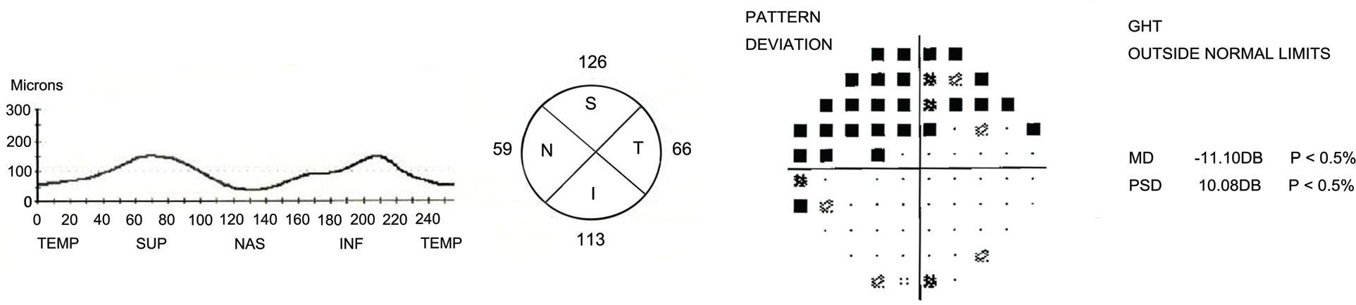
Figure 2.
Retinal nerve fiber layer (RNFL) thickness for normal, glaucoma suspect, and glaucoma eyes in children after adjusted by refractive error.
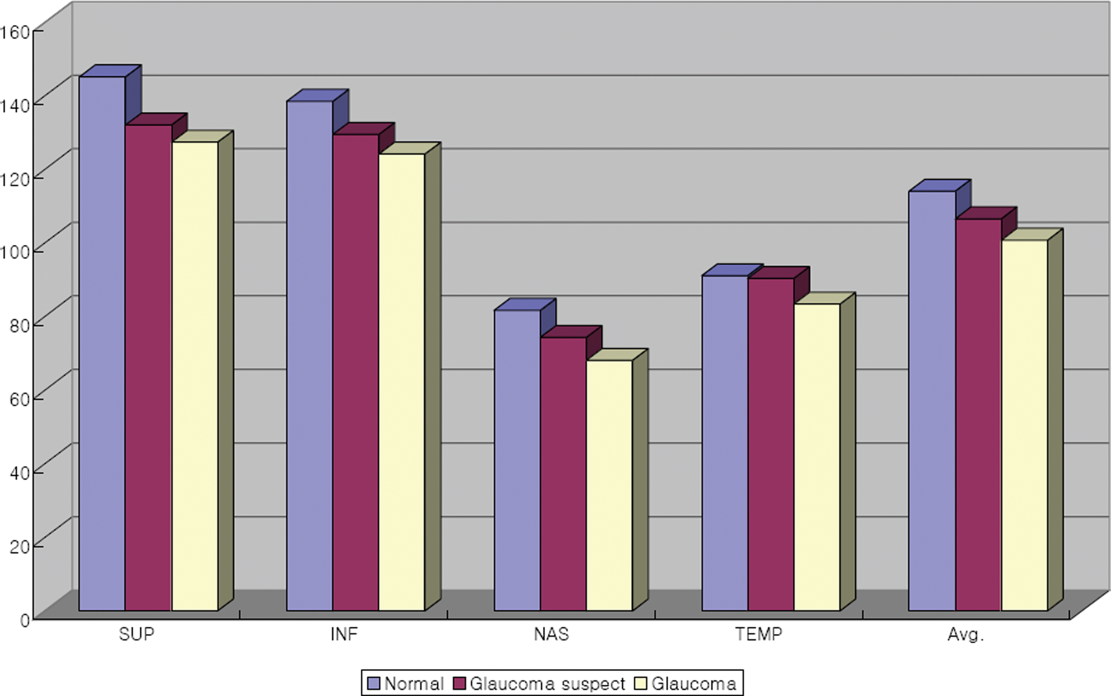
Table 1.
Demographics of the subjects’
| Normal | Glaucoma suspect | Glaucoma | p-value | ||
|---|---|---|---|---|---|
| Age (yr) | 12.5±3.97 | 13.2±3.24 | 12.9±4.19 | 0.182# | |
| Sex (n,eye) | male | 54 | 34 | 19 | 0.942∗∗ |
| Female | 40 | 28 | 14 | ||
| Ref. error (D)∗ | -1.54±0.97 | -2.41±1.13 | -2.53±1.52 | 0.016# | |
| Hyper. (n)† | 13 | 7 | 3 | ||
| Emetr. (n)‡ | 15 | 5 | 4 | 0.564∗∗ | |
| Myo. (n)§ | 66 | 50 | 26 | ||
| IOP (mmHg)∏ | 11.4±5.16 | 16.3±7.32 | 19.2±9.02 | <0.001# | |
| Max | 7 | 8 | 10 | ||
| Min | 20 | 28 | 29 |
Table 2.
Compositions of glaucoma suspect eyes in children
| n (eyes) P | Percentage (%) | |
|---|---|---|
| Total | 62 | 100 |
| Large CD∗ | 28 | 45.1 |
| Raised IOP† | 8 | 12.9 |
| Large CD∗+Medi.‡ | 6 | 9.7 |
| Raised IOP†+Medi.‡ | 10 | 16.1 |
| Large CD∗+Raised IOP†+Medi.‡ | 8 | 12.9 |
| CD∗ ratio asymmetry | 2 | 3.3 |
Table 3.
RNFL Thickness among three groups before adjusted by refractive error
| Location | Normal | p-value# | Glaucoma suspect | p-value∗∗ | Glaucoma | p-value†† |
|---|---|---|---|---|---|---|
| SUP (µm)∗ | 145.2±18.2 | 0.009 | 132.1±15.2 | 0.032 | 127.4±15.4 | 0.012 |
| INF (µm)† | 138.5±15.4 | 0.005 | 129.5±18.4 | 0.044 | 124.2±19.9 | 0.039 |
| NAS (µm)‡ | 81.7±15.2 | 0.013 | 74.4±16.8 | 0.022 | 68.1±19.2 | 0.015 |
| TEMP (µm)§ | 91.1±17.5 | 0.872 | 90.4±18.1 | 0.013 | 83.4±16.5 | 0.045 |
| Avg. Thickness (µm)∏ | 114.1±10.8 | 0.017 | 106.6±10.5 | 0.002 | 100.8±17.7 | <0.001 |
Table 4.
RNFL thickness among three groups after adjusted by refractive error
| Location | Normal | p-value# | Glaucoma suspect | p-value∗∗ | Glaucoma | p-value†† |
|---|---|---|---|---|---|---|
| SUP (µm)∗ | 140.1±17.5 | 0.021 | 134.8±15.5 | 0.037 | 129.5±16.5 | 0.030 |
| INF (µm)† | 134.1±14.8 | 0.026 | 130.2±18.8 | 0.048 | 126.1±20.2 | 0.047 |
| NAS (µm)‡ | 79.9±14.5 | 0.024 | 75.4±17.1 | 0.031 | 70.3±19.7 | 0.032 |
| TEMP (µm)§ | 89.2±16.8 | 0.902 | 91.2±18.7 | 0.017 | 85.1±16.9 | 0.064 |
| Avg. Thickness (µm)∏ | 110.8±10.1 | 0.039 | 107.9±10.8 | 0.011 | 102.8±18.1 | 0.003 |
 XML Download