

 PDF
PDF ePub
ePub Citation
 Print
Print


Abstract
Purpose
To evaluate the effectiveness and safety of treating orbital lesions with gamma knife radiosurgery (GKS).
Methods
Between April 2004 and January 2006, ten patients who had orbital tumors or vascular lesions and who underwent GKS were included in this retrospective study.
Results
Ten patients with orbital lesions were treated with GKS. The group of orbital lesions consisted of 5 meningiomas, 2 schwannomas, 1 cavernous hemangioma, 1 arteriovenous fistula, and 1 adenoidcystic carcinoma of the lacrimal gland. The most common symptom was proptosis. The tumors were located at the orbital apex in eight patients, and five of these patients were treated with fractionated stereotactic radiosurgery. The mean cumulative marginal dose was 17.0 Gy (12-20 Gy), and the mean cumulative maximal dose was 30.8 Gy (16.2–40.4 Gy). The follow-up period ranged from 3 to 25 months (mean 13.9 months). During the follow-up period, magnetic resonance imaging revealed a decrease of tumor volume in 3 patients with symptomatic improvement. In two patients, tumor volume increased. No radiation-induced optic neuropathy, retinopathy, or cataract was observed in any of the 10 patients during the follow-up period.
References
1. Maroon JC, Kennerdell JS. Surgical approaches to the orbit. Indications and techniques. J Neurosurg. 1984; 60:1226–35.
2. Lew H, Lee SY, Lee KS, et al. Removal of orbital tumor through transfrontal approach. J Korean Ophthalmol Soc. 1994; 35:1723–7.
3. Leksell L. Stereotactic radiosurgery. J Neurol Neurosurg Psychiatry. 1983; 46:797–803.

4. Leksell L. The stereotaxic method and radiosurgery of the brain. Acta Chir Scand. 1951; 102:316–9.
5. Pollock BE, Gorman DA, Schomberg PJ, Kline RW. The Mayo Clinic gamma knife experience: indications and initial results. Mayo Clin Proc. 1999; 74:5–13.
6. Yamamoto M. Gamma Knife radiosurgery: technology, applications, and future directions. Neurosurg Clin N Am. 1999; 10:181–202.
7. Aichholzer M, Bertalanffy A, Dietrich W, et al. Gamma knife radiosurgery of skull base meningiomas. Acta Neurochir (Wien). 2000; 142:647–52.
8. Chen JC, Giannotta SL, Yu C, et al. Radiosurgical management of benign cavernous sinus tumors: dose profiles and acute complications. Neurosurgery. 2001; 48:1022–30.
9. Feigl GC, Bonelli CM, Berghold A, Mokry M. Effects of gamma knife radiosurgery of pituitary adenomas on pituitary function. J Neurosurg. 2002; 97:415–21.
10. Kobayashi T, Kida Y, Mori Y. Gamma knife radiosurgery in the treatment of Cushing disease: long-term results. J Neurosurg. 2002; 97:422–8.
11. Pendl G, Schröttner O, Friehs GM, Feichtinger H. Stereotactic radiosurgery of skull base meningiomas. Stereotact Funct Neurosurg. 1995; 64:S11–8.
12. Sheehan JM, Vance ML, Sheehan JP, et al. Radiosurgery for Cushing's disease after failed transsphenoidal surgery. J Neurosurg. 2000; 93:738–42.
13. Tishler RB, Loeffler JS, Lunsford LD, et al. Tolerance of cranial nerves of the cavernous sinus to radiosurgery. Int J Radiat Oncol Biol Phys. 1993; 27:215–21.
14. Andrews DW, Faroozan R, Yang BP, et al. Fractionated stereotactic radiotherapy for the treatment of optic nerve sheath meningiomas: preliminary observations of 33 optic nerves in 30 patients with historical comparison to observation with or without prior surgery. Neurosurgery. 2002; 51:890–902.
15. Chang SD, Adler JR Jr. Treatment of cranial base meningiomas with linear accelerator radiosurgery. Neurosurgery. 1997; 41:1019–25.
16. Ove R, Kelman S, Amin PP, Chin LS. Preservation of visual fields after peri‐ sellar gamma‐ knife radiosurgery. Int J Cancer. 2000; 90:343–50.
17. Girkin CA, Comey CH, Lunsford LD, et al. Radiation optic neuropathy after stereotactic radiosurgery. Ophthalmology. 1997; 104:1634–43.
18. Leber KA, Bergloff J, Langmann G, et al. Radiation sensitivity of visual and oculomotor pathways. Stereotact Funct Neurosurg. 1995; 64:S233–8.
19. Movsas B, Movsas TZ, Steinberg SM, Okunieff P. Long-term visual changes following pituitary irradiation. Int J Radiat Oncol Biol Phys. 1995; 33:599–605.
20. Paek SH, Downes MB, Bednarz G, et al. Integration of surgery with fractionated stereotactic radiotherapy for treatment of nonfunctioning pituitary macroadenomas. Int J Radiat Oncol Biol Phys. 2005; 61:795–808.
21. Estrada J, Boronat M, Mielgo M, et al. The long-term outcome of pituitary irradiation after unsuccessful transsphenoidal surgery in Cushing's disease. N Engl J Med. 1997; 336:172–7.
22. Metellus P, Regis J, Muracciole X, et al. Evaluation of fractionated radiotherapy and gamma knife radiosurgery in cavernous sinus meningiomas: treatment strategy. Neurosurgery. 2005; 57:873–86.
23. Brada M, Ford D, Ashley S, et al. Risk of second brain tumour after conservative surgery and radiotherapy for pituitary adenoma. BMJ. 1992; 304:1343–6.
24. Nelson PB, Goodman ML, Flickenger JC, et al. Endocrine function in patients with large pituitary tumors treated with operative decompression and radiation therapy. Neurosurgery. 1989; 24:398–400.
25. Sachs RK, Brenner DJ. Solid tumor risks after high doses of ionizing radiation. Proc Natl Acad Sci U S A. 2005; 102:13040–5.
26. Shaw EG, Coffey RJ, Dinapoli RP. Neurotoxicity of Radiosurgery. Semin Radiat Oncol. 1995; 5:235–45.
27. Hudgins PA, Newman NJ, Dillon WP, Hoffman JC Jr. Radiation-induced optic neuropathy: characteristic appearances on gadolinium-enhanced MR. AJNR Am J Neuroradiol. 1992; 13:235–8.
28. Jiang GL, Tucker SL, Guttenberger R, et al. Radiation-induced injury to the visual pathway. Radiother Oncol. 1994; 30:17–25.
29. Leber KA, Bergloff J, Pendl G. Dose-response tolerance of the visual pathways and cranial nerves of the cavernous sinus to stereotactic radiosurgery. J Neurosurg. 1998; 88:43–50.
Figure 1.
T1-weighted axial magnetic resonance image (A) and sagittal images (B) before radiosurgery in case 6 with cavernous hemangioma in the left orbital apex. Eighteen months after radiosurgery (C, D) the tumor volume is reduced.
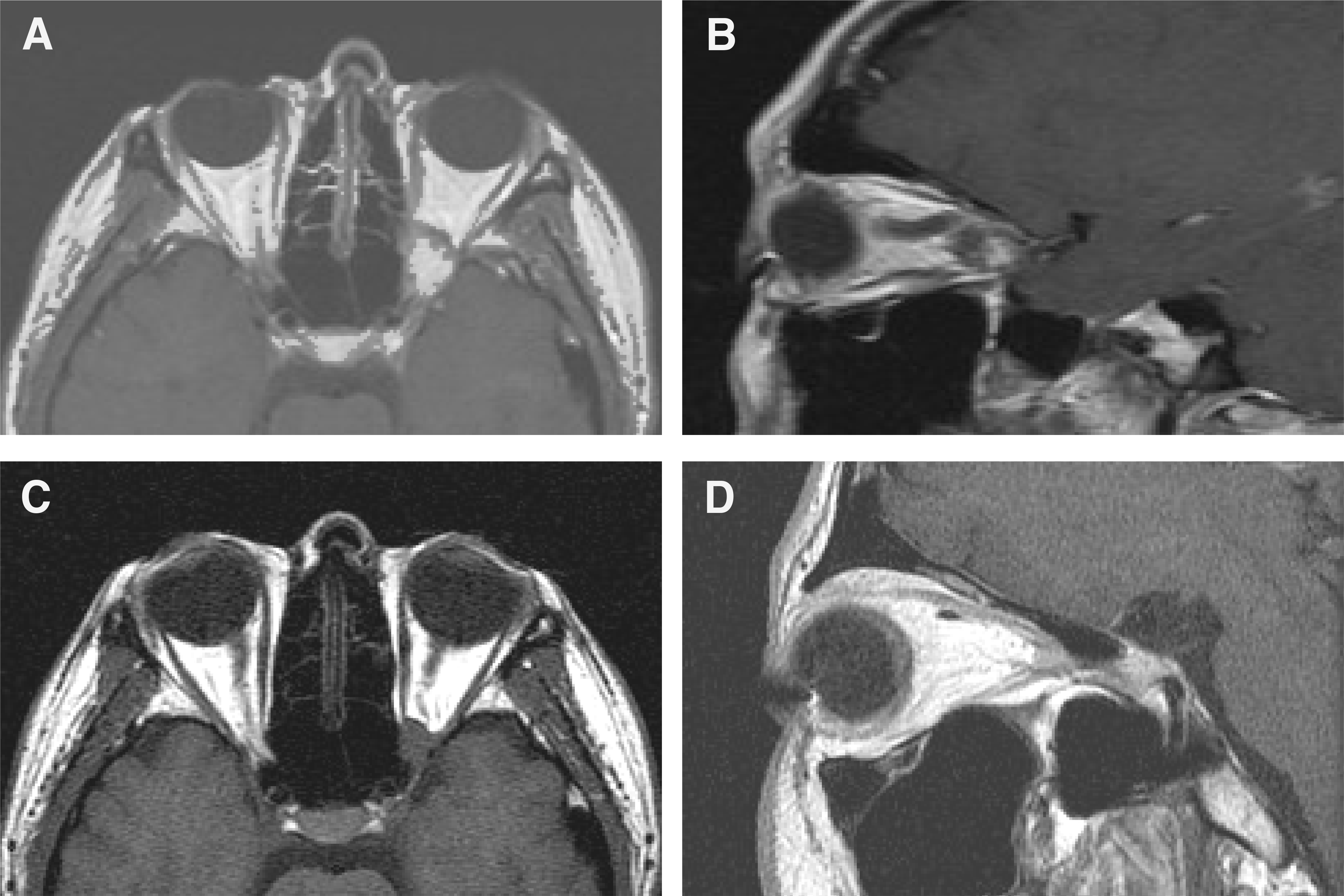
Figure 2.
Optic nerve sheath meningioma in a 34-year-old woman (case 1) presenting with decreased visual acuity in her left eye for 2 months. Before gamma knife surgery axial T1 contrast MR image (A) reveals a well-circumscribed retrobulbar mass in the left orbit and 7 months after fractionated gamma knife surgery (B) central necrotic lesion is shown. (C) Visual field examination of the left eye before radiosurgery showed extensive field loss. The visual acuity of her left eye decreased to 0.1. (D) Seven months after radiosurgery the visual field improved except small paracentral scotoma and visual acuity improved to 0.7.

Table 1.
Radiosurgical feature of lesions treated with gamma knife radiosurgery
| No. of patients | Pathologic diagnosis | Location | Maximal Dose (Gy) | Marginal dose (Gy) | No. of fraction |
|---|---|---|---|---|---|
| 1 | Meningioma | Optic n. | 10.2 | 5 | 3 |
| 2 | Meningioma | Optic n. cavernous sinus | 10.1 | 5 | 4 |
| 3 | Meningioma | Orbital apex | 25.5 | 12 | |
| 4 | Meningioma | Frontal & sphenoid bone | 8.0 | 4 | 4 |
| 5 | AVF* | Ophthalmic a. | 16.2 | 13 | |
| 6 | Cavernous hemangioma | Orbital apex | 31.5 | 15 | |
| 7 | Adenocystic ca. | Lacrimal gland | 26.2 | 13 | |
| 8 | Schwannoma | Orbital apex | 28.5 | 14 | |
| 9 | Meningioma | Optic n, | 10 | 5 | 4 |
| 10 | Schwannoma | Orbital apex | 9.1 | 5 | 4 |
Table 2.
Summary of results after radiosurgery
| No. of patients | Pathologic diagnosis | (months) Follow up |
Lesion Vol. (mm3) |
Radiosurgical |
Visual Acuity |
||
|---|---|---|---|---|---|---|---|
| Before GKS* | After GKS* | response | Before GKS* | After GKS* | |||
| 1 | Meningioma | 17 | 886.4 | 817.8 | Necrosis | 0.1 | 0.7 |
| 2 | Meningioma | 25 | 6800 | 4700 | Shrunken | LP†(+) | CF‡ |
| 3 | Meningioma | 13 | 2400 | 2074 | Static | 0.03 | 0.01 |
| 4 | Meningioma | 21 | 3100 | 8100 | Aggravated | 0.1 | LP (−) |
| 5 | AVF* | 16 | 32 | 25 | Static | 1.0 | 0.9 |
| 6 | Cavernous hemangioma | 18 | 2000 | 1400 | Shrunken | LP (−) | LP (−) |
| 7 | Adenocystic ca. | 3 | 2996.2 | 3879 | Aggravated | 1.0 | Exenteration |
| 8 | Schwannoma | 5 | 5000 | 4500 | Static | LP (+) | LP (+) |
| 9 | Meningioma | 9 | 736.6 | 733 | Static | 1.0 | 1.0 |
| 10 | Schwannoma | 12 | 1700 | 428 | Shrunken | 0.4 | 1.0 |
 XML Download