

 PDF
PDF ePub
ePub Citation
 Print
Print


Abstract
Purpose
To evaluate the distinguishing characteristics that may assist in the clinical diagnosis of sterile endophthalmitis from intravitreal triamcinolone injection.
Methods
From January 2005 to June 2006, the medical records of 163 eyes that received intravitreal triamcinolone injection were reviewed. In 5 eyes of 5 patients who complained of decreasing vision and mild ocular pain within three days after injection, slit lamp bimicroscopy revealed hypopyon, anterior chamber reaction, and vitritis. We analyzed clinical pictures retrospectively.
Results
Intravitreal triamcinolone injections were performed for diabetic macular edema in 4 patients and for panuveitis in 1 patient with Behçet's disease. Pseudophakia was found in all five eyes. Inflammations in three eyes resolved with topical and oral antibiotics within 13 days, and the other two patients were treated with a vitreous culture and intravitreal antibiotic injection. However, vitreous tap showed no evidence of endophthalmitis, and inflammation resolved within the same period.
Conclusions
Presumed sterile endophthalmitis presents within 3 days after intravitreal triamcinolone injection and may be accompanied by decreased vision and ocular discomfort, although it is characterized by no distinct ocular pain. The symptoms and ocular inflammation resolved quickly within 13 days with a favorable visual outcome. Inflammations in three eyes resolved without surgical intervention, so it may be necessary to closely observe clinical manifestations before assuming that the eye is suffering from infectious endophthalmitis.
References
1. Wilson CA, Berkowitz BA, Sato Y, et al. Treatment with intravitreal steroid reduces blood retinal barrier breakdown due to retinal photocoagulation. Arch Ophthalmol. 1992; 110:1155–9.
2. Martidis A, Duker JS, Greenberg PB, et al. Intravitreal triamcinolone for refractory diabetic macular edema. Ophthalmology. 2002; 109:920–7.

3. Jonas JB, Kreissig I, Sofker A, Degenring RF. Intravitreal triamcinolone for diffuse diabetic macular edema. Arch Ophthalmol. 2003; 121:57–61.
4. Greenberg PB, Martidis A, Rogers AH, et al. Intravitreal triamcinolone acetonide for macular edema due to central retinal vein occlusion. Br J Ophthalmol. 2002; 86:247–8.
5. Antcliff RJ, Spalton DJ, Stanford MR, et al. Intravitreal triamcinolone for uveitis cystoid macular edema: an optical coherence tomography study. Ophthalmology. 2001; 108:765–72.
6. Conway MD, Canaskis C, Livir-Rallatos C, Peyman GA. Intravitreal triamcinolone acetonide for refractory chronic pseudophakic cystoid macular edema. J Cataract Refract Surg. 2003; 29:27–33.
7. Wang YS, Friedrichs U, Eichler W, et al. Inhibitory effect of triamcinolone acetonide on bFGF– induced migration and tube formation in choroidal microvascular endothelial cells. Grafes Arch Clin Exp Ophthalmol. 2002; 240:42–8.
8. Ciulla TA, Criswell MH, Danis RP, Hill TE. Intravitreal triamcinoloe acetonide inhibits choroidal neovascularization in a laser– treated rat model. Arch Ophthalmol. 2001; 119:399–404.
9. Danis RP, Ciulla TA, Pratt LM, Anliker W. Intravitreal triamcinolone acetonide in exudative age related macular degeneration. Retina. 2000; 20:244–50.
10. Moshfeghi DM, Lowder CY, Roth DM, Kaiser PK. Retinal and choroidal vascular occlusion after posterior sub– tenon triamcinolone injection. Am J Ophthalmol. 2002; 134:132–4.
11. Oh MJ, Choi KR, Lee SY, Lee JH. Clinical manifestations of intraocular pressure elevation after intravitreal injection of triamcinolone acetonide. J Korean Ophthalmol Soc. 2006; 47:1575–82.
12. Özkiris A, Erkilic K. Complications of intravitreal injection of triamcinolone acetonide. Can J Ophthalmol. 2005; 40:63–8.
13. Cekic O, Chang S, Tseng JJ, et al. Cataract progression after intravitreal triamcinolone injection. Am J Ophthalmol. 2005; 139:993–8.
14. Jonas JB, Kreissig I, Degenring RF. Endophthalmitis after intravitreal injection of triamcinolone acetonide. Arch Ophthalmol. 2003; 121:1663–4.
15. Moshfehgi DM, Kaiser PK, Scott IU, et al. Acute endophthalmitis following intravitreal triamcinolone acetonide injection. Am J Ophthalmol. 2003; 136:791–6.
16. Nelson ML, Tennant MT, Sivalingam A, et al. Infectious and presumed noninfectious endophalmitis after intravitreal triamcinolone acetonide injection. Retina. 2003; 23:686–91.
17. Morrison VL, Koh HJ, Cheng L, et al. Intravitreal toxicity of the kenalog vehicle (benzyl alcohol) in rabbits. Retina. 2006; 26:339–44.
18. Hida T, Chandler D, Arena JE, Machemer R. Experimental and clinical observations of the intraocular toxicity of commercial corticosteroid preparation. Am J Ophthalmol. 1986; 101:190–5.
19. Narayanan R, Mungcal JK, Kenney MC, et al. Toxicity of triamcinolone acetonide on retinal neurosensory and pigment epithelial cells. Invest Ophthalmol Vis Sci. 2006; 47:722–8.
20. Jonas JB, Kreissig I, Spandau UH, et al. Infectious and noninfectious endophthalmitis after intravitreal high– dose triamcinolone acetonide. Am J Ophthalmol. 2006; 141:579–80.
21. Westfall AC, Osborn A, Kuhl D, et al. Acute endophthalmitis incidence: intravitreal triamcinolone. Arch Ophthalmol. 2005; 123:1075–7.
22. Wang LC. Yang CM. Sterile endophthalmitis following intravitreal injection of triamcinolone acetonide. Ocul Immunol Inflamm. 2005; 13:295–300.
23. Roth DB, Chieh J, Spirin MJ, et al. Noninfectious endophthalmitis associated with intravitreal triamcinolone injection. Arch Ophthalmol. 2003; 121:1279–82.
24. Sutter FK, Gilles MC. Pseudo– endophthalmitis after intravitreal injection of triamcinolone. Br J Ophthalmol. 2003; 87:972–4.
25. Moshfeghi DM, Kaiser PK, Scott IU, et al. Acute endophthalmitis following intravitreal triamcinolone acetonide injection. Am J Ophthalmol. 2003; 136:791–6.
26. Park HJ, Park JM, Oum BS. Presumed noninfectious endophthalmitis after intravitreal inejection of triamcinolone acetondie. J Korean Ophthalmol Soc. 2005; 46:1419–23.
Figure 1.
Anterior segment photograph. Multiple subconjunctival hemorrhage is observed at the site of incision. Hypopyon (<1 mm) in the anterior chamber (arrow) is seen on slit lamp examination.
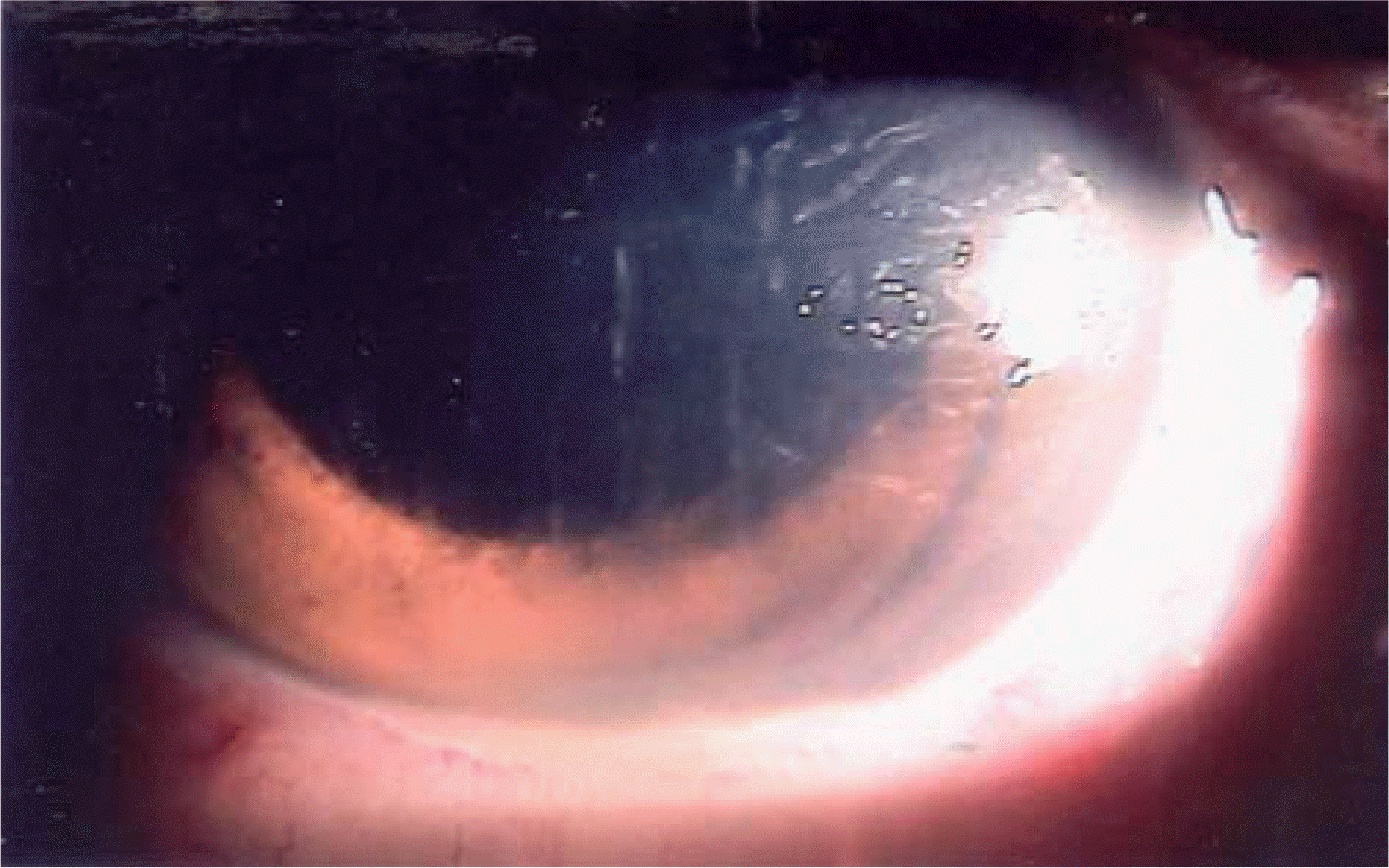
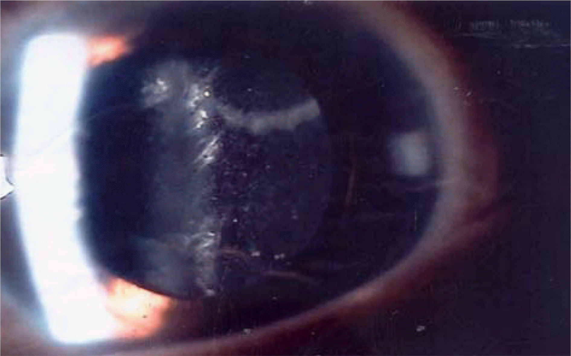
Figure 2.
Anterior segment photograph. The slit lamp examination shows triamcinolone strand (arrow) with vitreous haze in patient 5.
 XML Download