

 PDF
PDF ePub
ePub Citation
 Print
Print


Abstract
Purpose
To investigate the influence of preservation of an epithelial sheet in Epi-LASIK on postoperative pain and epithelial wound healing time.
Methods
This prospective study included 34 eyes of 17 patients with myopia who received Epi-LASIK. An epithelial flap was created using the epikeratome (Centurion SES, Norwood Abbey EyeCare, Australia). After the stroma was ablated using the MEL 80 (Carl Zeiss Meditec. Germany) excimer laser, the epithelial sheet was replaced on the stromal bed in one randomly selected eye of each patient, and removed in the contralateral eye. The pain scores at postoperative day 0, 1, 2, 5 and the numbers of days for the complete epithelial wound healing were compared between the sheet-preserved and the sheet-removed eyes. At 1 month postoperative, uncorrected visual acuity (UCVA), refractive error and corneal haze were also compared.
Results
Pain scores on the day of operation were lower in sheet-preserved group and statistically significant ( p=0.01). On postoperative day 1, 2, and 5, pain scores did not reach statistical significance ( p=0.24, 0.08, 0.56, respectively). The mean epithelial healing time was 4.88±0.93 days for the flap-preserved eyes and 4.29±0.77 days for the flap-removed eyes, which showed statistical significance ( p=0.01). No significant difference was noted between the 2 groups for mean UCVA, corneal haze and refractive error at 1 month postoperative.
References
1. Melki SA, Azar DT. LASIK complications: etiology, management, and prevention. Surv Ophthalmol. 2001; 46:95–116.
2. Alió JL, Artola A, Claramonte PJ. . Complications of photorefractive keratectomy for myopia: two year follow-up of 3000 cases. J Cataract Refract Surg. 1998; 24:619–26.

3. Loewenstein A, Lipshitz I, Varssano D, Lazar M. Complications of excimer laser photorefractive keratectomy for myopia. J Cataract Refract Surg. 1997; 23:1174–6.
4. Kim SY, Sah WJ, Lim YW, Hahn TW. Twenty percent alcohol toxicity on rabbit corneal epithelial cells; electron microscopic study. Cornea. 2002; 21:388–92.
5. Pallikaris IG, Kalyvianaki MI, Katsanevaki VJ, Ginis HS. Epi-LASIK: preliminary clinical results of an alternative surface ablation procedure. J Cataract Refract Surg. 2005; 31:879–85.
6. Pallikaris IG, Naoumidi II, Kalyvianaki MI, Katsanevaki VJ. Epi-LASIK: comparative histological evaluation of mechanical and alcohol-assisted epithelial separation. J Cataract Refract Surg. 2003; 29:1496–501.
7. O`Doherty M, Kirwan C, O`Keeffe M, O`Doherty J. Postoperative pain following Epi-LASIK, LASEK, and PRK for myopia J Refract Surg. 2007; 23:133–8.
8. Torres LF, Sancho C, Tan B. . Early postoperative pain following Epi-LASIK and Photorefractive Keratectomy: a prospective, comparative, bilateral study. J Refract Surg. 2007; 23:126–32.
9. Wilson SE, Liu JJ, Mohan RR. Stromal-epithelial interactions in the cornea. Prog Retin Eye Res. 1999; 18:293–309.
10. Imanishi J. Expression of cytokines in bacterial and viral infections and their biochemical aspects. J Biochem. 2000; 127:525–30.
11. Wilson SE, Mohan RR, Mohan RR. . The corneal wound healing response: cytokine-mediated interaction of the epithelium, stroma, and inflammatory cells. Prog Retin Eye Res. 2001; 20:625–37.
12. Gan L, Hamberg-Nystrom H, Fagerholm P, Van Setten G. Cellular proliferation and leukocyte infiltration in the rabbit cornea after photorefractive keratectomy. Acta Ophthalmol Scand. 2001; 79:488–92.
13. Andresen JL, Ehelers N. Chemotaxis of human keratocytes is increased by platelet-derived growth factor-BB, epidermal growth factor, transforming growth factor-alpha, acidic fibroblast growth factor, insulin-like growth factor-I, and transforming growth factor-beta. Curr Eye Res. 1998; 17:79–87.
14. Helena MC, Baerveldt F, Kim WJ. . Keratocyte apoptosis after corneal surgery. Invest Ophthalmol Vis Sci. 1998; 39:276–83.
15. Mohan RR, Hutcheon AE, Choi R. . Apoptosis, necrosis, proliferation, and myofibroblast generation in the stroma following LASIK and PRK. Exp Eye Res. 2003; 76:71–87.
16. Hamberg-Nystrom H, Baldwin HC, Dreiss AK. . Temporal relationships of the epithelial-stromal interaction in corneal wound healing ARVO abstract 3624. Invest Ophthalmol Vis Sci. 2000; 41:S681.
17. Wilson SE. Molecular cell biology for the refractive corneal surgeon: programmed cell death and wound healing. J Refract Surg. 1997; 13:171–5.
18. Wilson SE. Analysis of the keratocyte apoptosis, keratocyte proliferation, and myofibroblast transformation responses after photorefractive keratectomy and laser in situ keratomileusis. Trans Am Ophthalmol Soc. 2002; 100:411–33.
19. Gavrieli Y, Sherman Y, Ben-Sasson SA. Identification of programmed cell death in situ via specific labeling of nuclear DNA fragmentation. J Cell Biol. 1992; 119:493–501.
20. Wilson SE. Keratocyte apoptosis in refractive surgery: Everett Kinsey Lecture. CLAO J. 1998; 24:181–5.
21. Gao J, Gelber-Schwalb TA, Addeo JV, Stern ME. Apoptosis in the rabbit cornea after photorefractive keratectomy. Cornea. 1997; 16:200–8.
22. Piatigorsky J. Review: a case for corneal crystallins. J Ocul Pharmacol Ther. 2000; 16:173–80.
23. Jester JV, Moller-Pedersen T, Huang J. . The cellular basis of corneal transparency: evidence for corneal crystallins. J Cell Sci. 1999; 112:613–22.
24. Katsanevaki VJ, Naoumidi II, Kalyvianaki MI, Pallikaris G. Epi-LASIK: Histological findngs of separated epithelial sheets 24 hours after treatment. J Refract Surg. 2006; 22:151–4.
25. Tanioka H, Hieda O, Kawasaki S. . Assessment of epithelial integrity and cell viability in epithelial flaps prepared with the epi-LASIK procedure. J Cataract Refract Surg. 2007; 33:1195–200.
26. Lee HK, Lee KS, Kim JK. . Epithelial healing and clinical outcomes in excimer laser photorefractive surgery following three epithelial removal techniques: mechanical, alcohol, and excimer laser. Am J Ophthalmol. 2005; 139:56–63.
27. Szaflik JP, Ambroziak AM, Szaflik J. Therapeutic use of a lotrafilcon A silicone hydrogel soft contact lens as a bandage after LASEK surgery. Eye Contact Lens. 2004; 30:59–62.
28. Nishi Y, Nishi O, Nishi K, Auffarth G. Pain reduction after epi-LASIK with a simple surgical procedure J Cataract Refract Surg. 2007; 33:555–7.
Figure 1
. Case (A) and (B) represent the clinical appearance of the corneal epithelium 4 and 3 days after Epi-LASIK respectively. Loose epithelium or erosion-like lesion (arrow) and epithelial defect (arrowhead) were seen on slit lamp examination.

Figure 2
. Comparison of the mean epithelial healing rate between epithelial flap-preserved group and epithelial flap- removed group. * Wilcoxon signed ranks test p<0.05.
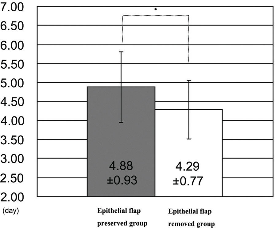
Table 1.
Preoperative characteristics of patients (Mean±SD)
| Epithelial flap-preserved group | Epithelial flap-removed group | p-value∗ | |
|---|---|---|---|
| Age (years) | 25.9±4.75 | ±4.75 | |
| BCVA†(logMAR) | -0.04±0.04 | -0.04±0.04 | 1.00 |
| S.E‡(diopter) | -4.72±1.72 | -4.43±1.53 | 0.09 |
Table 2.
Data of 11-point numeric pain scores after Epi-LASIK
Table 3.
Comparison of 11-point numeric pain scores between epithelial sheet preserved and removed group (Median pain score (range))
| Time after surgery | OP day | 1 day | 2 day | 5 day |
|---|---|---|---|---|
| Epithelial flap-preserved group | 4 (2-9) | 2 (1-5) | 2 (1-4) | 0 (0-1) |
| Epithelial flap-removed group | 5 (2-9) | 2 (1-6) | 2 (1-4) | 0 (0-1) |
| p-value∗ | 0.01 | 0.24 | 0.08 | 0.56 |
Table 4.
Comparison of UCVA and S.E after Epi-LASIK (Mean±SD)
| UCVA† (logMAR) after Epi-LASIK | S.E‡ (Diopter) | |||
|---|---|---|---|---|
| 1 week | 2 weeks | 1 month | 1 month | |
| Epithelial flap-preserved group | 0.11±0.10 | 0.03±0.04 | 0.02±0.04 | -0.05±0.60 |
| Epithelial flap-removed group | 0.10±0.08 | 0.05±0.05 | 0.02±0.04 | -0.11±0.33 |
| p-value∗ | 0.78 | 0.30 | 0.73 | 0.97 |
 XML Download