

 PDF
PDF ePub
ePub Citation
 Print
Print


Abstract
Purpose
To compare corneal topographic changes using Orbscn II between keratoconus and keratoconus-suspected eyes.
Methods
Thirty-seven keratoconus eyes, 17 keratoconus-suspected eyes and 37 normal eyes were evaluated by using Orbscan II corneal topography. We compared central phachymetry, anterior elevation from best-fit sphere (BFS), posterior elevation from BFS, most protruded corneal thickness, central corneal thickness, anterior chamber depth, corneal diameter, and pupil size.
Results
Central pachymetry, anterior and posterior elevation from BFS, central corneal thickness, and anterior chamber depth were statistically significantly different between keratoconus and control eyes. Anterior elevation from BFS showed a significant difference between keratoconus-suspected and control eyes. There were statistically significant differences in central pachymetry, posterior elevation from BFS, central corneal thickness and most protruded corneal thickness between keratoconus and keratoconus-suspected eyes. Corneal diameter and pupil size showed no differences among the 3 groups.
Conclusions
Suspected keratoconus eyes have a higher value of anterior elevation from BFS on Orbscan II topography as compared with control eyes. Central pachymetry, posterior elevation from BFS, central corneal thickness and most protruded corneal thickness may be helpful in distinguishing between keratoconus and keratoconus-suspected eyes.
References
1. Rabinowitz YS. Keratoconus. Surv Ophthalmol. 1998; 42:297–319.

2. Krachmer JH, Feder RS, Belin MW. Keratoconus and related noninflammatory corneal thinning disorders. Surv Ophthalmol. 1984; 28:293–322.
3. Caroline PJ, Doughman DJ, McGuire JR. Preliminary report on a new contact lens design for keratoconus. Contact Intraocul Lens. 1978; 4:69–73.
4. Soper JW, Jarrett A. Results of a systematic approach to fitting keratoconus and corneal transplants. Cont Lens Med Bull. 1972; 5:50–9.
5. Edrington TB, Barr JT, Zadnik K, et al. Standardized rigid contact lens fitting protocol for keratoconus. Optom Vis Sci. 1996; 73:369–75.
6. Rosenthal P. The Boston Lens and the management of keratoconus. Int Ophthalmol Clin. 1986; 26:101–9.
7. Maguire LJ, Bourne WM. Corneal topography of early keratoconus. Am J Ophthalmol. 1989; 108:107–12.
8. Maguire LJ, Lowry JC. Identifying progression of subclinical keratoconus by serial topography analysis. Am J Ophthalmol. 1991; 112:41–5.
9. Rao SN, Raviv T, Majmudar PA, Epstein RJ. Role of Orbscan II in screening keratoconus suspects before refractive corneal surgery. Ophthalmology. 2002; 109:1642–6.
10. Rabsilber TM, Becker KA, Frisch IB, Auffarth GU. Anterior chamber depth in relation to refractive status measured with the Orbscan II Topography System. J Cataract Refract Surg. 2003; 29:2115–21.
11. Pflugfelder SC, Liu Z, Feuer W, Verm A. Corneal thickness indices discriminate between keratoconus and contact lens induced corneal thinning. Ophthalmology. 2002; 109:2336–41.
12. Amsler M. The “forme fruste” of keratoconus. Wien klin Wocherschr. 1961; 73:842–3.
13. Wilson SE, Klyce SD. Advances in the analysis of corneal topography. Surv Ophthalmol. 1991; 35:269–77.
14. Guarnieri FA, Guarnieri JC. Comparison of Placido-based, rasterstereography, and slit-scan corneal topography system. J Refract Surg. 2002; 18:169–76.
15. Maeda N, Klyce SD, Smolek MK. Comparison of methods for detecting keratoconus using videokeratography. Arch Ophthalmol. 1995; 113:870–4.
16. Rabinowitz YS, McDonnel PJ. Computer-assisted corneal topography in keratoconus. Refract Corneal Surg. 1989; 5:400–8.
17. Rabinowitz YS. Videokeratographic indices to aid in screening for keratoconus. J Refract Surg. 1995; 11:371–9.
18. Rabinowitz YS, Rasheed K. KISA index: a quantitative videokeratography algorithm embodying minimal topographic criteria for diagnosing keratoconus. J Cataract Refract Surg. 2000; 26:472–4.
19. Smolek MK, Klyce SD. Current keratoconus detection methods compared with a neural network approach. Invest Ophthalmol Vis Sci. 1997; 38:2290–9.
20. Lee HH, Shin MC, Lee HB. The Analysis of Corneal Topography after Laser in Situ Keratomileusis (LASIK) for Myopic Correction. J Korean Ophthalmol Soc. 2001; 42:960–6.
21. Liu Z, Huang AJ, Pflugfelder SC. Evaluation of corneal thickness and topography in normal eyes using the Orbscan corneal topography system. Br J Ophthalmol. 1999; 83:774–8.
22. Kim JD, Park KS, Kim SD. Corneal thickness variation and consistency according to daytime. J Korean Ophthalmol Soc. 2000; 41:1690–6.
23. Choi HJ, Kim MK, Lee JL. Diagnostic criteria for keratoconus using Orbscan II slit scanning topography/pachymetry system. J Korean Ophthalmol Soc. 2004; 45:928–35.
24. Auffarth GU, Wang L, Volcker HE. Keratoconus evaluation using the Orbscan Topography System. J Cataract Refract Surg. 2000; 26:222–8.
25. Kawana K, Miuta K, Tokunaga T, et al. Central corneal thickness measurements using Orbscan II scanning slit topography, noncontact specular microscopy, and ultrasonic pachymetry in eyes with keratoconus. Cornea. 2005; 24:967–71.
26. Arntz A, Duran JA, Pijoan JI. Subclinical keratoconus diagnosis by elevation topography. Arch Soc Esp Oftalmol. 2003; 78:659–64.
27. Mohammad HD, Hassan H. A quantitative corneal topography index for detection of keratoconus. J Refract Surg. 1998; 14:427–36.
Figure 1.
The map of Orbscan II topography in keratoconus. The parameters including central corneal power, anterior and posterior elevation from BFS and central corneal thickness are significantly different from normal eyes.
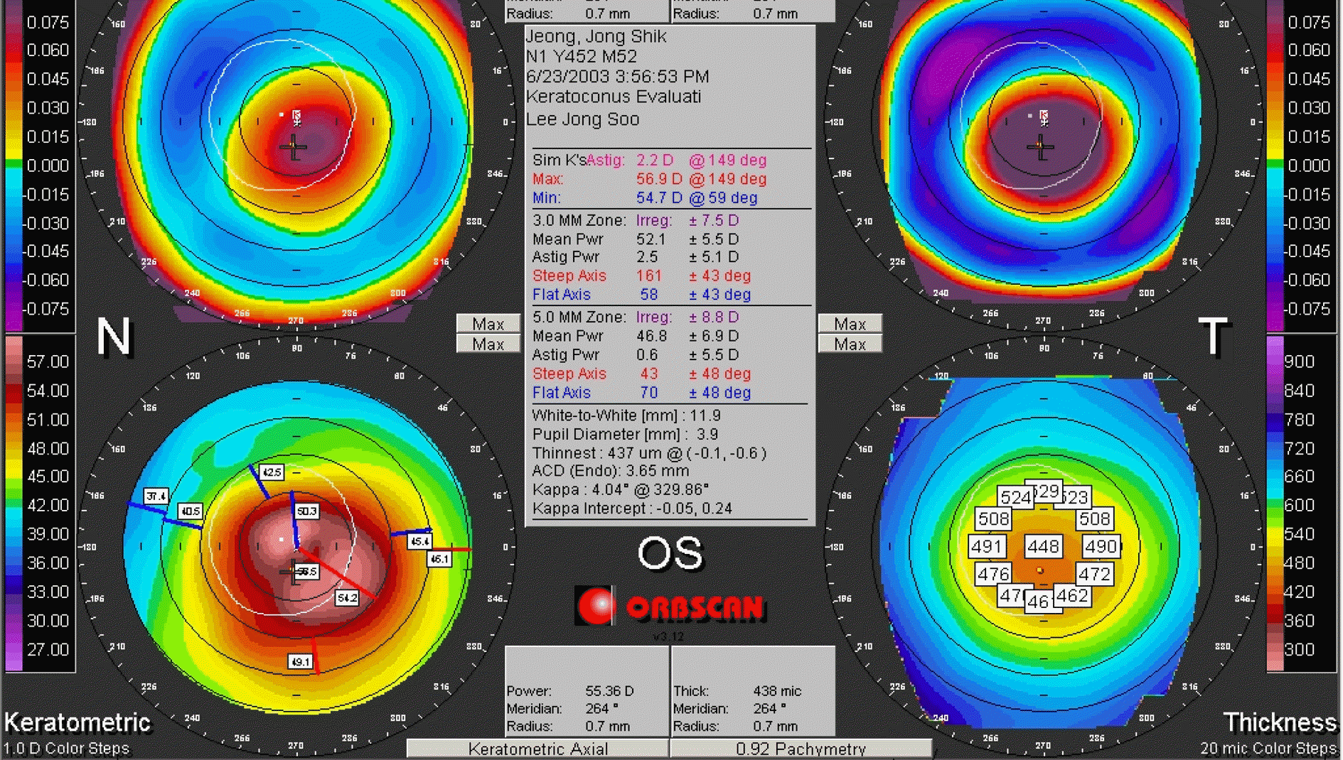
Figure 2.
The map of Orbscan II topography in keratoconus suspect. Anterior elevation from BFS is the only significantly different parameter comparing with normal eyes, and central corneal power, posterior elevation from BFS, central corneal thickness and most protruded corneal thickness are the parameters significantly different from keratoconic eyes.
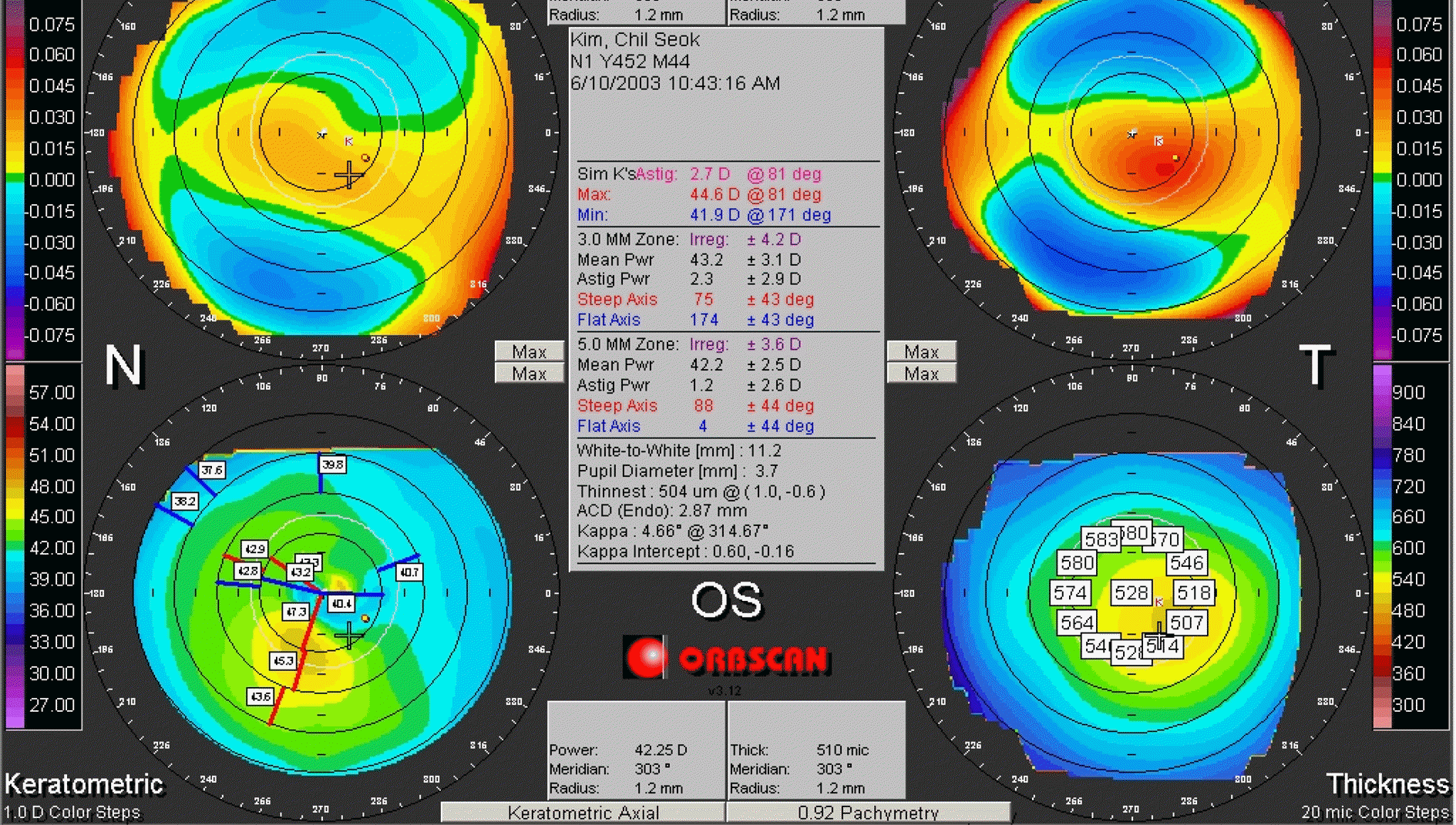
Table 1.
Comparison of parameters of Orbscan II system in keratoconic and normal eyes. The parameters including central corneal power, anterior and posterior elevation from BFS and central corneal thickness are significantly different from normal eyes
| Parameter | Keratoconic eye | Normal eye | p-value |
|---|---|---|---|
| Central corneal power (Diopter) | 48.81±6.33 | 42.48± 1.71 | < 0.001 |
| (Range) | (37.60∼72.06) | (38.98∼44.81) | |
| Anterior elevation from BFS* (μm) | 43.05±27.26 | 6.80±4.91 | < 0.001 |
| (Range) | (4.00∼125.00) | (1.00∼14.00) | |
| Posterior elevation from BFS* (μm) | 93.93±54.64 | 25.70±7.13 | < 0.001 |
| (Range) | (9.00∼225.00) | (15.00∼41.00) | |
| Central corneal thickness (pm) | 451.07±61.01 | 543.65±44.24 | < 0.001 |
| (Range) | (282.00∼560.00) | (441.00∼611.00) | |
| Anterior chamber depth (mm) | 3.34±0.36 | 3.07±0.23 | 0.024 |
| (Range) | (2.64∼4.04) | (2.73∼3.47) | |
| Corneal diameter (mm) | 11.74±0.36 | 11.56±0.45 | 0.104 |
| (Range) | (11.10∼12.50) | (11.10∼13.00) | |
| Pupil size (mm) | 4.91±1.48 | 5.15± 1.44 | 0.564 |
| (Range) | (3.20∼10.80) | (3.70∼8.00) |
Table 2.
Comparison of parameters of Orbscan II system in keratoconus-suspected and normal eyes. Anterior elevation from BFS is the only significantly different parameter compared with normal eyes
| Parameter | Keratoconus-suspected eye | Normal eye | p-value |
|---|---|---|---|
| Central corneal power (Diopter) | 42.74± 1.59 | 42.48± 1.71 | 0.999 |
| (Range) | (40.32∼46.12) | (38.98∼44.81) | |
| Anterior elevation from BFS* (μm) | 24.28±7.65 | 6.80±4.91 | 0.014 |
| (Range) | (6.00∼37.00) | (1.00∼14.00) | |
| Posterior elevation from BFS* (μm) | 30.72±19.03 | 25.70±7.13 | 0.591 |
| (Range) | (1.00∼64.00) | (15.00∼41.00) | |
| Central corneal thickness (pm) | 501.84±55.34 | 543.65±44.24 | 0.065 |
| (Range) | (362.00∼580.00) | (441.00∼611.00) | |
| Anterior chamber depth (mm) | 3.27±0.35 | 3.07±0.23 | 0.188 |
| (Range) | (2.45∼3.83) | (2.73∼3.47) | |
| Corneal diameter (mm) | 11.81±0.43 | 11.56±0.45 | 0.101 |
| (Range) | (10.80∼12.50) | (11.10∼13.00) | |
| Pupil size (mm) | 4.95±1.24 | 5.15±1.44 | 0.662 |
| (Range) | (3.40∼7.80) | (3.70∼8.00) |
Table 3.
Comparison of parameters of Orbscan II system in keratoconic and keratoconus-suspected eyes. Central corneal power, posterior elevation from BFS, central corneal thickness and most protruded corneal thickness are the parameters significantly differed from keratoconus-suspected eyes
| Parameter | Keratoconic eye | Keratoconus suspected eye | p-value |
|---|---|---|---|
| Central corneal power (Diopter) | 48.81±6.33 | 42.74±1.59 | < 0.001 |
| (Range) | (37.60∼72.06) | (40.32∼46.12) | |
| Anterior elevation from BFS* (μm) | 43.05±27.26 | 24.28±7.65 | 0.980 |
| (Range) | (4.00∼125.00) | (6.00∼37.00) | |
| Posterior elevation from BFS* (μm) | 93.93±54.64 | 30.72±19.03 | < 0.001 |
| (Range) | (9.00∼225.00) | (1.00∼64.00) | |
| Central corneal thickness (μm) | 451.07±61.01 | 501.84±55.34 | 0.003 |
| (Range) | (282.00∼560.00) | (362.00∼580.00) | |
| Most protruded corneal thickness (μm) | 440.05±66.38 | 497.38±59.13 | 0.001 |
| (Range) | (280.00∼560.00) | (362.00∼579.00) | |
| Anterior chamber depth (mm) | 3.34±0.36 | 3.27±0.35 | 0.845 |
| (Range) | (2.64∼4.04) | (2.45∼3.83) | |
| Corneal diameter (mm) | 11.74±0.36 | 11.81±0.43 | 0.563 |
| (Range) | (11.10∼12.50) | (10.80∼12.50) | |
| Pupil size (mm) | 4.91±1.48 | 4.95± 1.24 | 0.924 |
| (Range) | (3.20∼10.80) | (3.40∼7.80) |
 XML Download