

 PDF
PDF ePub
ePub Citation
 Print
Print


Abstract
Purpose
To evaluate the factors that affect the eyelid height changes during the postoperative period in patients who underwent levator resection under local anesthesia.
Methods
Among the 242 patients that underwent levator resection under local anesthesia by the same surgeon between January on 1995 and December 2003, marginal reflex distance 1 (MRD1) measurements were performed using a caliper in 91 patients who were followed for more than 3 months.
Results
There were 36 males and 55 females, aged between 12 and 78 years (average of 33.6 years). The average follow-up period of the patients was 8.7 months (3 months ∼ 58 months). During this period, 86 patients (94.5%) experienced satisfactory results. The average change in the MRD1 of the eyelids preoperatively, during the operation, and 1 week, 1 month, 3 months, 6 months, and 1 year postoperatively were 0.8 mm, 3.9 mm, 3.0 mm, 2.7 mm, 2.5 mm, 2.4 mm, and 2.2 mm, respectively. The MRD1 decreased 1.2 mm after 1 month and stabilized. When the levator function was greater than 8 mm, the height of the eyelids stabilized within 1 week. The worse the function of the levator palpebrae, such as in the case of congenital ptosis, the greater the correction was needed.
References
1. Anderson RL, Dixon RS. Aponeurotic ptosis surgery. Arch Ophthalmol. 1979; 97:1123–8.

2. Older JJ. Levator aponeurosis surgery for the correction of acquired ptosis : analysis of 113 procedures. Ophthalmology. 1983; 90:1056–9.
3. Lee JY. A statistical study on the corneal diameters in Korean. J Korean Ophthalmol Soc. 1983; 24:53–8.
4. Linberg JV, Vasquez RJ, Chao GM. Aponeurotic ptosis repair under local anesthesia : prediction of results from operative lid height. Ophthalmology. 1988; 95:1046–52.
5. Crawford JS. Frontalis sling operation. J Pediatr Ophthalmol Strabismus. 1982; 19:253–5.
6. Lee KY, Chi YH, Woo KI, Kim YD. Frontalis sling operation using preserved fascia lata. J Korean Ophthalmol Soc. 1997; 38:2084–90.
7. Chung PJ, Chung WS. Aponeurotic ptosis surgery. J Korean Ophthalmol Soc. 1990; 31:255–63.
8. Mauriello JA, Wagner RS, Caputo AR, et al. Treatment of congenital ptosis by maximal levator resection. Ophthalmology. 1985; 93:466–9.
9. Fasanella RM, Servat J. Levator resection for minimal ptosis : another simplified operation. Arch Ophthalmol. 1961; 65:493–6.
10. Choe KS, Kim YS, Lee TS. A clinical study of surgical results on 456 blepharoptosis. J Korean Ophthalmol Soc. 1995; 36:1093–104.
11. Anderson RL, Beard C. The levator aponeurosis : Attachments and their clinical significance. Arch Ophthalmol. 1977; 95:1437–41.
12. Jones LT. The anatomy of the upper eyelid and its relation to ptosis surgery. Am J Ophthamol. 1964; 57:943–59.
13. Kim YD, Shin KS. Surgical correction of acquired ptosis due to an aponeurotic defect. J Korean Ophthalmol Soc. 1990; 31:837–44.
14. Linberg JV, Vasquez RJ, Chao GM. Aponeurotic ptosis repair under local anesthesia : prediction of results from operative lid height. Ophthalmology. 1988; 95:1046–52.
15. Yang HS, Ahn JH, Kim SJ, Han YS. Surgical results of levator resection using the MLD in congenital blepharoptosis. J Korean Ophthalmol Soc. 2000; 41:2247–53.
Figure 1.
Measurements of marginal reflex distance 1 (MRDi) and horizontal corneal diameter from each photograph using a caliper.
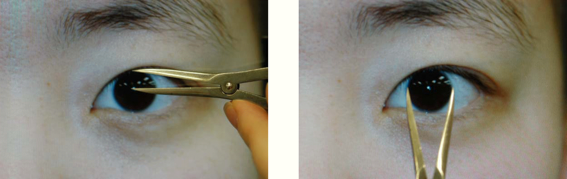
Figure 3.
(A) Preoperative photograph of a unilateral congenital ptosis patient. (B) One year after unilateral levator resection under local anesthesia.
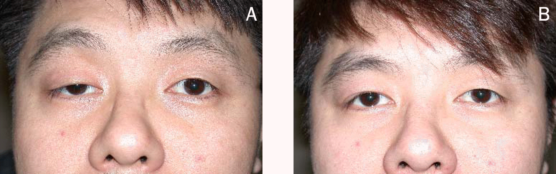
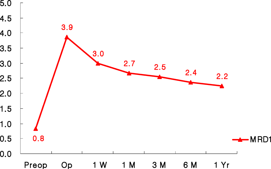
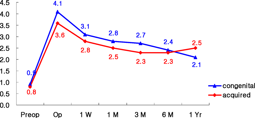
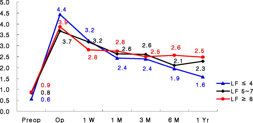
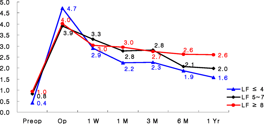
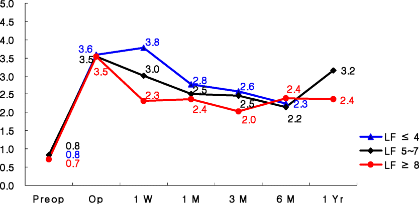
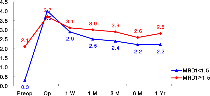
Table 1.
Number of patients in the age groups by sex and laterality
Table 2.
Classification of ptosis patients
| Classification | Number of patients (%) |
|---|---|
| Congenital ptosis | 56 (61.5%) |
| simple | 56 |
| Acquired ptosis | 35 (38.5%) |
| involutional | 23 |
| traumatic | 12 |
 XML Download