

 PDF
PDF ePub
ePub Citation
 Print
Print


INTRODUCTION
Mandibular third molar extraction is one of the most commonly performed procedures in dentistry. Most patients' primary concern is pain during dental extraction. Pain control is an essential part of dental extraction, and the most common method of pain control is local anesthesia.
Local anesthesia may be categorized in a number of ways according to the extent of the area to be anesthetized. To anaesthetize the mandible, there are many local anesthesia methods that target the inferior alveolar nerve, which runs along the mandibular canal. The conventional inferior alveolar nerve block has been used frequently in various procedures for many years. However, the success rate of the inferior alveolar nerve block is, in fact, only modest, and associated complications, such as aspiration and nerve injury, are fairly common. Thus, various anesthesia methods have been continuously studied to try to address this issue [1]. In addition, while lidocaine has been the preferred anesthetic for local anesthesia in Korea, in recent years, the use of articaine, which is known to have a higher success rate than lidocaine in achieving anesthesia, is also increasing. [23]
In this article, we review, with current updates, the various methods of inferior alveolar nerve anesthesia, local anesthetics used in these methods, and the computer-controlled local anesthetic delivery system (CCLAD), which has been known to provide nearly painless delivery of local anesthesia, for mandibular third molar extraction.
LOCATION OF THE MANDIBULAR FORAMEN
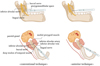
In order to increase the success rate of anesthesia, the location of the mandibular foramen (MF) must be accurately known. As it is not possible to palpate the MF, surrounding anatomical landmarks, such as the occlusal plane, sigmoid notch, coronoid notch, and external and internal oblique ridges are used [4]. Thangavelu et al. showed that the MF is not positioned at the midpoint of anteroposterior width of the ramus, but rather situated at a distance 2.75 mm posterior to the midpoint of the ramus and approximately 19 mm from the coronoid notch. They also found that the MF is positioned at the level of or slightly below the occlusal plane and is situated approximately 3mm above the midpoint of the imaginary line running from the sigmoid notch and the inferior border of the mandible [5]. Narayana et al. showed that the MFs of three year olds, nine year olds, and adults were positioned at 4.12 mm below, near the level of, and 4.16 mm above the occlusal plane, respectively [6]. Most studies have shown that in adults, the MF was, in general, situated at the level of or below the occlusal plane. As the nerves move about 4mm posteriorly when the mouth is opened, needle insertion of 23 mm in length is needed with respect to the coronoid notch on the occlusal plane [5].
INFERIOR ALVEOLAR NERVE BLOCK TECHNIQUES
1. Conventional inferior alveolar nerve block
The conventional inferior alveolar nerve block is the most commonly used nerve block technique in dentistry. This technique involves anesthesia of the inferior alveolar nerve, which enters the mandibular foramen, via the positioning of the needle on the mandibular foramen area. The crucial clinical landmarks of this technique are the coronoid notch and pterygomandibular raphe. The insertion point is located 3/4 down the line drawn from the deepest part of the pterygomandibular raphe to the coronoid notch. The needle must be advanced until the bone is contacted. Aspiration is mandatory prior to administration of the local anesthetics and administration should be done very slowly.
The nerves anesthetized are the inferior alveolar, incisor, mental, and lingual nerves. The mandibular teeth to the midline, the body of the mandible, the lower part of the mandibular ramus, buccal periosteum and mucous membrane to the premolars, anterior 2/3 of the tongue, oral floor, lingual soft tissue, and the periosteum are all anesthetized [1]. The failure rate of the conventional inferior alveolar nerve block is greater than 20%. Anatomical variations of the mandible and insufficient insertion depth into the soft tissue are thought to be the key factors in nerve block failure [1].
2. Gow-gates technique
In 1973, George Albert Edwards Gow-Gates proposed a new technique [7]. This technique has a higher success rate than the conventional inferior alveolar nerve block [8]. It has a blood aspiration rate of approximately 2%, which is lower when compared to that of the conventional inferior alveolar nerve block (10~15%) [9]. In this block technique, the needle is positioned just inferior to the mesiolingual cusp of the upper second molar and advanced slowly until it makes bony contact with the frontal side of the condylar. As the insertion height of this technique is higher on the occlusal plane of the mandible than that of the conventional inferior alveolar nerve block, the Gow-Gates technique anesthetizes the inferior alveolar, mental, incisor, lingual, mylohyoid, auriculotemoporal, and buccal nerves in about 75% of patients [10].
The areas anesthetized are the mandibular teeth to the midline, buccal mucoperiosteum, mucous membranes of the insertion area, anterior 2/3 of the tongue, oral floor, lingual soft tissue, periosteum, body of the mandible, lower part of the mandibular ramus, skin of the zygomatic bone, and the posterior side of buccal and temporal areas [1]. The advantages of the Gow-Gates technique include less pain during insertion when compared to the conventional inferior alveolar nerve block [11] and anesthetization of a more extensive area with a single injection. A disadvantage of the Gow-Gates technique is the slower onset of anesthesia when compared to the conventional inferior alveolar nerve block [1].
3. Arched needle technique
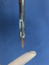
According to the data reported by Ashish Chakranarayan, the failure rate may be lowered if the needle is arched at an angle almost perpendicular to the ramus and inserted to the medial side of the ramus. The needle is inserted slightly posterior to that in the conventional technique and to 4–5 mm in depth parallel to the occlusal plane. Then, the embedded tip is used as a pivot to arch the uninserted portion of the needle posteromedially, so that the approaching angle of the needle tip is changed from acute to nearly perpendicular [12].
Although the pre-bending of needles is against safe practices due to the risk of needle breakage and tissue tear [13], the authors have checked the safety of the fine 27 gauge needles used in the study by bending them prior to insertion. (Fig. 1 and 2). A success rate of 98% has been reported for this technique [12].
4. Mandibular foramen anterior technique
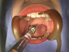
The conventional inferior alveolar nerve block has potential risks including neural or vascular injury. Takasugi et al. introduced a technique that can eliminate such risks. As opposed to the conventional technique, in which the needle tip is directed at the mandibular foramen, this technique positions the needle anterior to the mandibular foramen [14]. According to a radiographic study by Okamoto et al., the anesthetic solution rapidly reached the mandibular foramen when the anesthetic was injected anteriorly to the mandibular foramen, thereby providing experimental support for this anesthesia technique [15]. In this technique, the needle tip is positioned at a distance approximately 10 mm above the occlusal plane of the mandible and inserted to a depth of 10 mm towards the lateral side of the pterygomandibular raphe from the contralateral first molar, creating an approaching angle of 60.1 ± 7.1°, which is greater compared to that of the conventional technique (49.7 ± 5.3°) (Fig. 3). The success rate was 75%. Although the success rate of this technique is not significantly different from that of the conventional technique, the advantage of the anterior technique is that it can reduce the risk of nerve injury or blood aspiration [14].
5. Fischer 1-2-3 technique
The Fischer 1-2-3 technique, which is also called the indirect technique, requires the identification of several anatomical landmarks, including the internal and external oblique ridge, and coronoid notch. In this technique, the anesthetic is injected using three different needle positions.
To achieve buccal anesthesia, the needle is first positioned on the midpoint of the thumbnail when the thumb is placed on the external oblique from over the contralateral premolars and inserted to a depth of about 6 mm. Then, the needle is pulled out and moved to the same side so that the needle slides onto the internal oblique ridge. The syringe is maintained parallel to the mandible occlusal plane and the needle is advanced about 8 mm. Then, the syringe is repositioned over the opposite first premolar and the needle is advanced 12–15 mm until the tip makes contact with bone. The needle should be withdrawn a bit and aspirated before the anesthetic is injected. [161718].
6. New technique introduced by Boonsiriseth et al.
Boonsiriseth et al. have introduced a technique for achieving anesthesia of the inferior alveolar nerve without periosteum contact. The insertion point is the same as that of the conventional inferior alveolar nerve block and the syringe is positioned parallel to the mandibular occlusal plane of the same side of the surgical site. The insertion depth is controlled by a rubber stop. A 30 mm needle is used and, with the rubber stop positioned at 20 mm, the needle is advanced until the rubber stop makes contact. As the needle does not contact the periosteum, this technique provides less pain compared to the conventional technique and reduces the frequency of positive aspiration and the risk of neural or vascular injury [1920].
7. Technique proposed by Thangavelu et al.
The conventional technique requires a number of anatomical landmarks and has a high failure rate due to indistinct anatomical structures. Thangavelu et al., proposed an anesthesia technique that uses the internal oblique ridge as the only anatomical landmark. When the thumb is placed on the retromolar area, the internal oblique ridge is indicated by the tip of the thumb. The insertion point will be 6–8 mm above the midpoint of the thumb and 2 mm posterior to the internal oblique ridge. The syringe is placed over the contralateral premolars and the needle is advanced until it touches the bone. The success rate of this anesthesia technique was 95% [1720].
INFERIOR ALVEOLAR NERVE BLOCK DRUGS
1. Lidocaine
Lidocaine was the first amide type local anesthetic to be was synthesized. Compared to the ester type local anesthetics, lidocaine has an extremely low rate of allergic reactions, and provides a fast onset of anesthesia, and a relatively long duration of anesthesia, making it the most commonly used local anesthetic in dentistry. For dental procedures, 2% lidocaine with 1:100,000 epinephrine is typically used, and for procedures in which bleeding may be a problem, 1: 50,000 epinephrine is recommended for hemostasis. The maximum recommended dose of epinephrine is 0.2mg for healthy adults. When using a 1.8 mL cartridge, approximately 11 cartridges may be used. For patients with cardiovascular dysfunction, the maximum recommended dose is 0.04mg, which is approximately equal to the amount contained in two cartridges. Lidocaine overdosing can lead to initial drowsiness followed by loss of consciousness and respiratory failure later on [1]. In order to prolong the duration of anesthesia, highly concentrated lidocaine may be used. However, caution is advised as the toxicity also increases [21]. Deo reported that a supplemental, submucosal injection of dexamethasone at the insertion site of the inferior alveolar nerve block prolongs the duration of anesthesia [22].
2. Articaine
Articaine is the only amide type local anesthetic to contain an ester group. It is typically used in 4% concentration. Its duration of action is similar to those of other local anesthetics and must be used with vasoconstrictors due to its vasodilation effect. The thiophene ring structure of articaine increases lipid solubility, enabling it to diffuse more easily when compared to the other local anesthetics [1]. In addition, the ester group of articaine is hydrolyzed by plasma esterase, and therefore has shorter half-life than other amide type anesthetics, thus reducing the risk of toxicity from overdosing [23].
It has been reported that the duration of the articaine anesthetic effect was longer when 2% lidocaine and 4% articaine were used for the inferior alveolar nerve block [2]. Moreover, the number of cases in which re-anesthesia was needed was lower in the articaine group [3]. In other words, the anesthesia success rate when using 4% articaine was higher than when 2% lidocaine was used. It should be noted, however, that a higher incidence of paresthesia has been reported in association with the use of 4% articaine compared to other anesthetics in some studies [24]. It has also been reported that when achieving nerve blockage using articaine, the high diffusibility of articaine may lead to ophthalmologic complications [25]. When the same concentration of articaine (4%) and lidocaine (4%) were used, articaine demonstrated a more rapid onset of anesthesia. Pain evaluations showed that the 4% lidocaine group experienced less pain than the 4% articaine group, though the difference was not statistically significant [26].
CCLAD (COMPUTER-CONTROLLED LOCAL ANESTHETIC DELIVERY)
Computer-controlled local anesthetic delivery (CCLAD) system was first introduced in dentistry in 1997. This computerized system automatically controls the injection speed and dose of local anesthetics, allowing uniform injection from one injection to another. In other words, the use of such a device reduces pain by continuously administering a small amount of anesthetic at a slow rate [1].
CCLAD devices are designed differently depending on whether the anesthetic cartridge is included in the main unit, the speed and mode of drug injection, aspiration function, and weight. If the anesthetic cartridge is included in the main unit, the hand piece is light and easier to use. When using the CCLAD system, the operator must hold the hand-piece stationary during administration of anesthesia, and therefore, a hand-piece that is too heavy causes difficulties with the maintenance of a stationary grip, which then increase the risk of needle movement into the tissue and needle breakage [27].
The Wand (Milestone Scientific, USA) is the most widely known product and consists of a main unit, foot pedal, and hand-piece. The single-use, disposable hand-piece is lightweight and provides great tactile sensation. Moreover, the hand-piece is held in a pen-like grasp, allowing the operator to easily rotate the needle after insertion. It is typically used for the hard palate, attached gingiva, and periodontal ligament anesthesia. However, it can also provide more accurate anesthetic delivery when deep tissue anesthesia, such as an inferior alveolar nerve block, is needed. In addition, both injection speed and pressure can be precisely controlled for all types of local anesthetics, allowing consistent pressure and speed of injection to be maintained even in areas with low flexibility [128].
The Midwest Comfort Control Syringe (Dentsply, USA) does not include a foot pedal and has a two-level delivery system. The injection begins at a very slow rate and starts to inject at a set rate after 10 seconds. It has five pre-programmed speeds, which can be set according to the anesthetic technique being used. The advantage of this system is that it allows the operator to select various injection rates. The disadvantage of this system is that it is bulkier than other devices and therefore difficult to use [27]. Other CCLAD devices have also been introduced in recent years and are being widely used.
DISCUSSION
The conventional alveolar nerve block is, in general, the most widely used local anesthesia technique for mandibular third molar extraction. However, the failure rate of this technique is high, and, depending on the cases, this nerve block often fails, even when performed by an experienced operator [13]. Failures of the conventional technique may be partly attributed to anatomical factors. You et al. reported that the success rate of anesthesia of the retrognathic group was lower than that of the other groups. They suggested that this may be related to the relatively shorter condylar length in patients with a retrognathic mandible, in which case the position of the mandibular foramen is higher than in normal mandibles. If the anesthetic is injected at a higher point than normal in patients with retrognathic mandibles, the success rate of anesthesia may be increased [29]. Although the Gow-Gates technique has a higher success rate than the conventional technique and does not require an additional insertion for buccal nerve anesthesia, the delayed onset of action and difficulty of mastering the technique have been pointed out as its limitations [1]. In the arched needle technique, proposed by Ashish Chakranarayan, the safety of the needle is checked by bending the needle prior to use as there is a possibility of needle breakage after insertion. This practice however seems to increase the risk of mechanical needle breakage and its practical clinical use is deemed difficult. The mandibular foramen anterior technique can reduce the incidence of blood aspiration and risk of nerve injury; however, its success rate is relatively low, at 75%, and further studies are needed to increase the success rate.
An inferior alveolar nerve block may cause restricted jaw opening, paralysis of the lingual nerve, paresthesia, dysesthesia, and, in rare cases, ophthalmologic complications [1]. In contrast, infiltration anesthesia has relatively a short duration of anesthesia without the complications of a nerve block. Moreover, filtration anesthesia is less painful during injection and does not come with the risk of tongue or lip numbness. Due to these advantages, many clinicians have been performing infiltration anesthesia using articaine and there has been an increasing interest in its use [30]. In contrast to other amide type local anesthetics, articaine has been shown to be highly infiltrative to the cancellous bone of the mandible. Nonetheless, it is believed the varying success rates reported of infiltration anesthesia using articaine in mandibular third molar extraction [3031] are due to various mandibular cortical bone thicknesses. Flanagan reported that the cortical bone thickness of 2–3 mm is the cutoff point for successful anesthesia in mandible posterior infiltration anesthesia, suggesting the possibility of employing infiltration anesthesia using articaine alone to achieve anesthesia [32]. In other words, infiltration anesthesia may provide sufficient anesthesia in dental extractions for patients with a thin mandibular cortical bone. A high possibility of inferior alveolar or lingual nerve injury after a nerve block using articaine has been reported in some studies; however, the authors would like to report that, up to now, they have not encountered any particular problems using articaine to achieve nerve blockage.
 XML Download