

 PDF
PDF ePub
ePub Citation
 Print
Print


Endotracheal intubation is an essential procedure to prevent patients from aspirating blood [1] and facilitate clear visualization of the surgical field during oral and maxillofacial surgery. However, various intraoperative complications associated with the use of this tube, such as tube obstruction, kinking, and dislodgement; leakage; and accidental extubation, have been reported [2]. In particular, direct tube damage has been reported to occur during intraoral surgery [34]. Here we describe a case of damage to the pilot balloon of a nasotracheal tube during orthognathic double-jaw surgery in a 27-year-old man. The damaged tube was cut and exchanged for a new one through an airway exchange catheter.
CASE REPORT
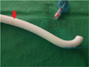
A 27-year-old man (168 cm, 65 kg) with no specific medical history was hospitalized to undergo elective high Le Fort I osteotomy and bilateral sagittal split osteotomy under general anesthesia for mandibular prognathism and midfacial deficiency. His preoperative laboratory findings, chest radiograph, and electrocardiogram were normal. He received intramuscular glycopyrrolate as premedication. His baseline blood pressure, heart rate, and oxygen saturation were 125/78 mmHg, 67 beats/min, and 99%, respectively. General anesthesia was induced by propofol 120 mg and rocuronium 60 mg. Following adequate muscle relaxation, nasotracheal intubation was attempted using a north facing nasal endotracheal tube (Portex® tube; Smiths Medical International, Hythe, UK) with an internal diameter of 6.5 mm. We checked the condition of the tube before induction and found no cuff leakage. After intubation, the patient was mechanically ventilated with 6 vol% desflurane and 3 L/min of fresh gas flow with 50% oxygen in air. The ventilator settings were as follows: tidal volume (VT), 500 mL; frequency, 10/min. The end tidal concentration of carbon dioxide (EtCO2) was 30 mmHg and the airway pressure was 18 cmH2O. Approximately 3 h into the surgery, in the middle of maxillary osteotomy using a reciprocating saw and osteotome, the surgeon was alerted about air bubble formation in the oral cavity blood. Furthermore, VT and minute ventilation had decreased to 350 mL and 4 L, respectively. Gap formation between the trachea and tube cuff was initially suspected; accordingly, air was injected using a syringe through the pilot balloon. However, repeated ballooning was ineffective. Immediately after the ballooning attempt, pilot balloon shrinkage was observed, leading to a suspicion of tube or cuff damage. The pilot balloon had not completely collapsed; a low tube pressure (10 mmHg) was detected by an endotracheal tube cuff pressure gauge. Continuation of surgery without exchanging the damaged tube was deemed impossible. Therefore, the anesthesiologist entered the surgical field after wearing an aseptic gown and gloves. Ventilation with 100% oxygen and suction was initiated. The portion of the tube outside the nostril was cut and an aseptic and lubricated airway exchange catheter (Cook airway exchange catheter, Cook critical care, USA) was inserted to remove the damaged tube. A new nasotracheal tube was immediately inserted through the airway exchange catheter. Examination of the damaged tube revealed an intact cuff; however, there was a small, superficial cut in the center of the pilot balloon (Fig. 1). Surgery was continued and successfully completed 2 h later. Nasotracheal intubation was maintained because of airway edema and bleeding. The patient's postoperative course was uneventful without any complications.
DISCUSSION
Endotracheal intubation is performed for airway maintenance, prevention of aspiration of gastric contents, and mechanical ventilation [5]. Most of the complications associated with this procedure develop during intubation and extubation, while complications caused by the surgery itself are rare. Roelofse et al. [6] reported a case of nasotracheal tube perforation by a Kirschner wire that was being used for fixation of a maxillary fracture. Another report documented tracheal tube cuff perforation caused by central catheterization via the subclavian vein during ventriculoatrial shunting [7].
Damage to the nasotracheal tube is more common during oral and maxillofacial surgery than during other surgeries because the surgical field, which is the intraoral space, is close to the tube; moreover, such surgeries require the use of a saw and osteotome [8]. In the present case, a reciprocating saw and osteotome were used to separate the maxilla from the anterior nasal spine during Le Fort I osteotomy, and this was majorly responsible for the nasotracheal tube damage. In most other cases of endotracheal tube damage, wide severance of the tube was reported. However, the surgeon operating on the present case was confident that there was no extensive damage. Minute ventilation was barely sustained, indicating that the cuff had not completely collapsed, and examination of the retrieved tube revealed a small, superficial cut in the pilot balloon. Coincidentally, slight injury by saw or osteotome influenced on narrow pathway of pilot tube.
In case of intraoperative damage to the endotracheal tube, methods of tube exchange are available, including the use of a rigid laryngoscope, an airway exchange catheter, or a fiberoptic bronchoscope [910]. However, direct rigid laryngoscopic reintubation is difficult in oral surgeries because edema and blood in the surgical field may obstruct the field of vision. Fiberoptic nasotracheal intubation is also difficult because of edema in the contralateral nasal cavity. For the prevention of surgical site contamination, reintubation using an airway exchange catheter was a suitable method for our patient. Airway exchange catheters have a lumen for jet ventilation, which is advantageous and can be used to provide oxygen ventilation in emergencies [11].
In conclusion, the findings from this case suggest that nasotracheal tubes can be damaged more easily during the intraoperative period of oral and maxillofacial surgery because of close proximity with the surgical field, and such damage is associated with many significant complications. In the present case, a small injury impaired normal airway management. Therefore, anesthesiologists must be vigilant, monitor the patient carefully, and work closely with the surgeon. Furthermore, the surgeon must be careful and use protective equipment for the tube during surgery in order to avoid such complications.
 XML Download