

 PDF
PDF ePub
ePub Citation
 Print
Print


Abstract
Purpose
Few studies have investigated the neurological complications of rotavirus infection in newborns. This study reports on clinical characteristics of newborns with seizures during rotavirus infection period and the neurological complications through case reports of infants who experienced seizures during rotavirus infection and demonstrated deep white matter injury on brain magnetic resonance imaging (MRI).
Methods
The study retrospectively investigated the medical records of newborn patients with positive rotavirus results who admitted to the neonatal intensive care unit of Dong-a University Hospital between January 2010 and May 2015. We analyzed the clinical characteristics of patients with seizures compared to without seizures among the patients of positive rotavirus results. The clinical characteristics of seizure patients according to the stool rotavirus antigen test results (positive vs. negative) and MRI results (normal vs. abnormal) were compared and analyzed.
Results
Of the 144 infants with positive rotavirus results, 17 infants (11.8%) had the seizures. These 17 patients showed no other symptom except seizures, positive rotavirus results predominantly at 4-6 days after birth, and more abnormal MRI results compared to without seizures. These results investigated in the seizure infants with positive rotavirus and infants of abnormal MRI results with positive rotavirus in same trend. In 15 newborns who showed positive stool test results and abnormal MRI results, seizure symptoms occurred at 4-6 days after birth, mostly following a pattern of clonic seizures. On MRI, all these newborns showed cerebral deep white matter injury in areas including the corpus callosum (CC) and internal capsule. Six out of 15 newborns underwent follow-up MRI scans, and five of them showed porencephalic white matter disease or periventricular leukomalacia.
Figures and Tables
Fig. 1
Diffusion-weighted images of the patients. Diffusion-weighted images of the 3 patients (A; patient 8, B; patient 9, C; patient 10) of the group of positive rotavirus and abnormal MRI. Note the high signals on deep white matter, corpus callosum and internal capsule.
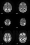
Fig. 2
Follow-up magnetic resonance image (MRI) after 2 weeks from seizure onset of patients A shows porencephaly and cerebromalacia in both frontal and parietal white matter, periventricular leukomalacia around occipital horn (A-1, A-2; patient 8). Likewise, porencephaly in frontal and parietal white matter is demonstrated in follow-up MRI of patient B (B-1, B-2; patient 9).
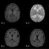
Table 1
Comparison of the Clinical Characteristics between Infants with Seizures and without Seizures in Rotavirus Group
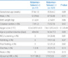
Table 2
Comparison of the Clinical Characteristics between Infants with Positive Rotavirus and Negative Rotavirus in Seizure Group
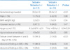
Table 3
Comparison of the Clinical Characteristics between Infants with Abnormal MRI and Normal MRI in Seizure Group
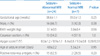
Table 4
Comparison of the Clinical Characteristics between Infants with Positive Rotavirus and Negative Rotavirus in Abnormal MRI Group
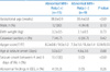
Table 5
Clinical Characteristics, Seizure Type, MRI and EEG Findings of the Group of Positive Rotavirus and Abnormal MRI
Abbreviations: GA, gestatioanal age; Bwt, birth weight; Del, delivery method; A/S, apgar score; DWI, diffusion-weighted imaging; EEG, electroencephalography; F, focal; M, multifocal; G, generalized; CS, corpus callosum; IC, internal capsule; EC, external capsule; T, thamus; BG, basal ganglia; NS, non-specific
References
1. Choi UY, Lee SY, Ma SH, Jang YT, Kim JY, Kim HM. Epidemiological changes in rotavirus gastroenteritis in children under 5 years of age after the introduction of rotavirus vaccines in Korea. Eur J Pediatr. 2013; 172:947–952.
2. Kim CR. Neonatal rotavirus infection. Neonatal Med. 2013; 20:389–401.

3. Park DK, Chung JY. The changes in the outbreak of rotavirus gastroenteritis in children after introduction of rotavirus vaccines: a retrospective study at a tertiary hospital. Korean J Pediatr Infect Dis. 2014; 21:167–173.
4. Sohn TY, Lee CJ, Kim YJ, Kang MJ, Kim SH, Lee SY, et al. Clinical and epidemiological study of 1,165 hospitalized cases of rotaviral gastroenteritis before and after the introduction of rotavirus vaccine, 2006-2013. Korean J Pediatr Infect Dis. 2014; 21:174–180.
5. Langley JM, LeBlanc JC, Hanakowski M, Goloubeva O. The role of Clostridium difficile and viruses as causes of nosocomial diarrhea in children. Infect Control Hosp Epidemiol. 2002; 23:660–664.
6. Gurwith M, Wenman W, Hinde D, Feltham S, Greenberg H. A prospective study of rotavirus infection in infants and young children. J Infect Dis. 1981; 144:218–224.
7. Santosham M, Yolken RH, Quiroz E, Dillman L, Oro G, Reeves WC, et al. Detection of rotavirus in respiratory secretions of children with pneumonia. J Pediatr. 1983; 103:583–585.
8. Park MK, Park JO, Kim CH. Comparison of clinical manifestation of rotaviral gastroenteritis between neonates and infants. Korean J Pediatr Gastroenterol Nutr. 2006; 9:153–161.
9. Lynch M, Lee B, Azimi P, Gentsch J, Glaser C, Gilliam S, et al. Rotavirus and central nervous system symptoms: cause or contaminant? Case reports and review. Clin Infect Dis. 2001; 33:932–938.
10. Shiihara T, Watanabe M, Honma A, Kato M, Morita Y, Ichiyama T, et al. Rotavirus associated acute encephalitis/encephalopathy and concurrent cerebellitis: report of two cases. Brain Dev. 2007; 29:670–673.
11. Lee KY, Oh KW, Weon YC, Choi SH. Neonatal seizures accompanied by diffuse cerebral white matter lesions on diffusion-weighted imaging are associated with rotavirus infection. Eur J Paediatr Neurol. 2014; 18:624–631.
12. Salmi TT, Arstila P, Koivikko A. Central nervous system involvement in patients with rotavirus gastroenteritis. Scand J Infect Dis. 1978; 10:29–31.
13. Riedel F, Kroener T, Stein K, Nuesslein TG, Rieger CH. Rotavirus infection and bradycardia-apnoea-episodes in the neonates. Eur J Pediatr. 1996; 155:36–40.
14. Verboon-Maciolek MA, Truttmann AC, Groenendaal F, Skranes J, Døllner H, Hunt RW, et al. Development of cystic periventricular leukomalacia in newborn infants after rotavirus Infection. J Pediatr. 2012; 160:165–168.
15. Wong CJ, Price Z, Bruckner DA. Aseptic meningitis in an infant with rotavirus gastroenteritis. Pediatr Infect Dis. 1984; 3:244–246.
16. Ushijima H, Bosu K, Abe T, Shinozaki T. Suspected rotavirus encephalitis. Arch Dis Child. 1986; 61:692–694.
17. Keidan I, Shif I, Keren G, Passwell JH. Rotavirus encephalopathy: evidence of central nervous system involvement during rotavirus infection. Pediatr Infect Dis J. 1992; 11:773–775.
 XML Download