Abstract
Purpose:
The aim of this study was to evaluate the result of extraarticular dorsal closing wedge osteotomy in Freiberg’s disease.
Materials and Methods:
Between February 2012 and July 2014, total 10 patients who underwent dorsal closing wedge osteotomy and followed up more than 1 year were selected for inclusion. Average age was 16.3 years, and average follow-up period was 15.5 months. The diagnosis was made using magnetic resonance imaging of those with a limitation in walking or usual activity due to pain in the metatarsal head. During operation, we removed loose body, and synovectomy was done. Osteotomy at the metatarsal neck and fixation with Kirschner wire were performed. X-ray was taken to check shortening of 2nd metatarsal and bone union. Moreover, we checked the active range of motion of 2nd metatarsophalangeal joint before and after surgery. At the last follow-up, the shortening of metatarsal, American Orthopaedic Foot and Ankle Society (AOFAS) score, visual analogue scale (VAS), and patient’s subjective satisfaction were evaluated.
Results:
According to the Smillie’s stage, there were 3 cases of stage II, 4 cases of stage III, and 3 cases of stage IV. Average bone union time on the osteotomy site was 8 weeks. Average shortening of metatarsal was 2.53 mm. Average AOFAS score improved significantly from 56.9 to 82.8 points at final follow-up (p<0.05), and average VAS score also improved significantly from 6.4 to 1.4 points at final follow-up (p<0.05). Average active range of motion at metatarsophalangeal joint improved from 28.0° preoperatively to 46.5° at the final follow-up. Other complications, such as metatarsalgia and arthritis, were not found; however, there was 1 case of delayed union with no symptom.
REFERENCES
1.Freiberg AH. Infraction of the second metatarsal bone: a typical injury. Surg Gynecol Obstet. 1914. 19:191–3.
2.Carmont MR., Rees RJ., Blundell CM. Current concepts review: Freiberg’s disease. Foot Ankle Int. 2009. 30:167–76.

31.Young K., Kim J., Joh J. Freiberg’s disease and metatarsophalan-geal joint instability. J Korean Foot Ankle Soc. 2013. 17:11–6.
4.Lee HS., Kwon SY., Kim DW., Chung JW. Weil osteotomy for Frei-berg’s disease. J Korean Foot Ankle Soc. 2011. 15:217–22.
5.Jones S., Al Hussainy HA., Ali F., Betts RP., Flowers MJ. Scarf osteotomy for hallux valgus. A prospective clinical and pedobaro-graphic study. J Bone Joint Surg Br. 2004. 86:832–6.
7.Al-Ashhab ME., Kandel WA., Rizk AS. A simple surgical technique for treatment of Freiberg’s disease. Foot (Edinb). 2013. 23:29–33.

8.Yoo CI., Jung CY., Kim BC., Choi SJ., Jung YW. Operative treatment of old neglected Freiberg’s infraction (comparison of three techniques). J Korean Foot Ankle Soc. 2004. 8:142–8.
9.Gauthier G., Elbaz R. Freiberg’s infraction: a subchondral bone fatigue fracture1 A new surgical treatment. Clin Orthop Relat Res. 1979. 142:93–5.
11.Yoon JH., Park SS., Kim EG., Lee CW. Treatment of Freiberg’s disease with joint debridement and reshaping of metatarsal head. J Korean Orthop Assoc. 1998. 33:1056–62.

12.Lee HJ., Kim JW., Min WK. Operative treatment of Freiberg disease using extra-articular dorsal closing-wedge osteotomy: technical tip and clinical outcomes in 13 patients. Foot Ankle Int. 2013. 34:111–6.
13.Kinnard P., Lirette R. Freiberg’s disease and dorsiflexion oste-otomy. J Bone Joint Surg Br. 1991. 73:864–5.

14.Chao KH., Lee CH., Lin LC. Surgery for symptomatic Freiberg’s disease: extraarticular dorsal closing-wedge osteotomy in 13 patients followed for 2-4 years. Acta Orthop Scand. 1999. 70:483–6.

15.Lee SK., Chung MS., Baek GH., Oh JH., Lee YH., Gong HS. Treatment of Freiberg disease with intra-articular dorsal wedge osteotomy and absorbable pin fixation. Foot Ankle Int. 2007. 28:43–8.

16.Kim J., Choi WJ., Park YJ., Lee JW. Modified Weil osteotomy for the treatment of Freiberg’s disease. Clin Orthop Surg. 2012. 4:300–6.

17.Stanley D., Betts RP., Rowley DI., Smith TW. Assessment of etiologic factors in the development of Freiberg’s disease. J Foot Surg. 1990. 29:444–7.
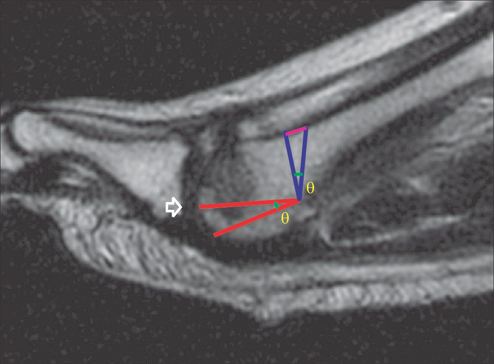
Figure 1.
Triangle was drawn at metatarsal neck to measure the amount of osteotomy using the tangent trigonometric ratio. Arrow, rotation of center.

Figure 2.
Plain radiographs and photographs of a 14-year-old female patient. (A, B) Preoperative anteroposterior (AP) and lateral show osteophytes and collapse of second metatarsal head and joint destruction. (C) Preoperative magnetic resonance imaging T2 proton density view show joint effusion and combination of low and high signal intensity with osteonecrosis. Postoperative AP radiograph (D) and last follow-up AP and lateral radiographs (E, F) show well maintained osteotomy site without joint destruction.

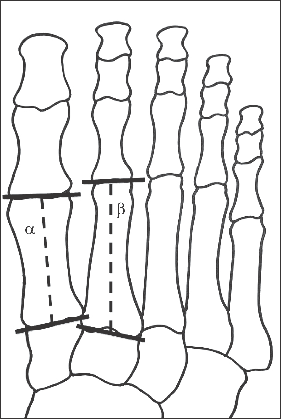
Figure 3.
Jone’s method that mesure the length of the second metatarsal. Line α and β are drawn along the longitudinal axis of the metatarsals. Metatarsal shortening=[{β preoperative × (α postoperative/α preoperative)}– β postoperative]

Table 1.
Dermographic Data of the Patients


 PDF
PDF ePub
ePub Citation
 Print
Print


 XML Download