

 PDF
PDF ePub
ePub Citation
 Print
Print


Abstract
Four patients with soft tissue defects around the ankle joint were covered with peroneal artery perforator-based propeller flaps. Using color Doppler sonography, the flap was designed by considering the location of the perforator and soft tissue defects. The procedure was then performed by rotating the flap by 180o. Additional skin graft was required in a patient due to partial necrosis, and delayed wound repair was performed in another patient with poor blood circulation at the distal part of the flap. The remaining patients did not have any complications and results were considered excellent. Good outcomes were eventually obtained for all patients.
REFERENCES
1.Rad AN., Singh NK., Rosson GD. Peroneal artery perforator-based propeller flap reconstruction of the lateral distal lower extremity after tumor extirpation: case report and literature review. Microsurgery. 2008. 28:663–70.

2.Schaverien M., Saint-Cyr M. Perforators of the lower leg: analysis of perforator locations and clinical application for pedicled per-forator flaps. Plast Reconstr Surg. 2008. 122:161–70.
3.Mateev MA., Kuokkanen HO. Reconstruction of soft tissue de-fects in the extremities with a pedicled perforator flap: series of 25 patients. J Plast Surg Hand Surg. 2012. 46:32–6.
4.Yoo MC., Chung DW., Han CS., Kim KH., Ahn JS. A clinical study of buoy flap. J Korean Orthop Assoc. 1987. 22:1157–65.
5.Chung DW., Hwang JS. Peroneal perforator flap. J Korean Soc Microsurg. 2004. 13:29–35.
6.Masquelet AC., Gilbert A., Restrepo J. The plantar flap in reconstructive surgery of the foot. Presse Med. 1984. 13:935–6.
7.Recalde Rocha JF., Gilbert A., Masquelet A., Yousif NJ., Sanger JR., Matloub HS. The anterior tibial artery flap: anatomic study and clinical application. Plast Reconstr Surg. 1987. 79:396–406.
8.Kneser U., Brockmann S., Leffler M., Haeberle L., Beier JP., Dragu A, et al. Comparison between distally based peroneus brevis and sural flaps for reconstruction of foot, ankle and distal lower leg: an analysis of donor-site morbidity and clinical outcome. J Plast Reconstr Aesthet Surg. 2011. 64:656–62.
9.Ensat F., Babl M., Conz C., Fichtl B., Herzog G., Spies M. Doppler sonography and colour Doppler sonography in the preoperative assessment of anterolateral thigh flap perforators. Handchir Mikrochir Plast Chir. 2011. 43:71–5.
Figure 1.
Dominant perforator is seen with blood flow towards the transducer (seen in red color) in this color Doppler sonography.
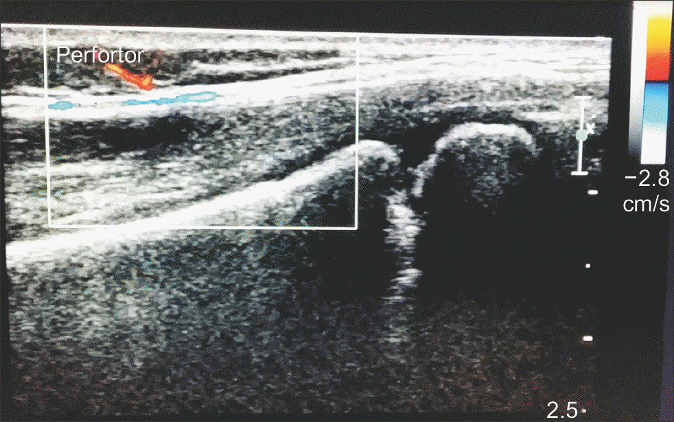
Figure 2.
These pictures show coverage of the posterior soft tissue defect with infected Achilles tendon using peroneal artery perforator propeller flap. (A) The perforator was identified and the flap was designed accordingly. (B) The flap was rotated with the perforator as a pivot. (C) The flap was inset and proximal wound was closed primarily. (D) This picture shows postoperative outcome at 7 months follow-up.
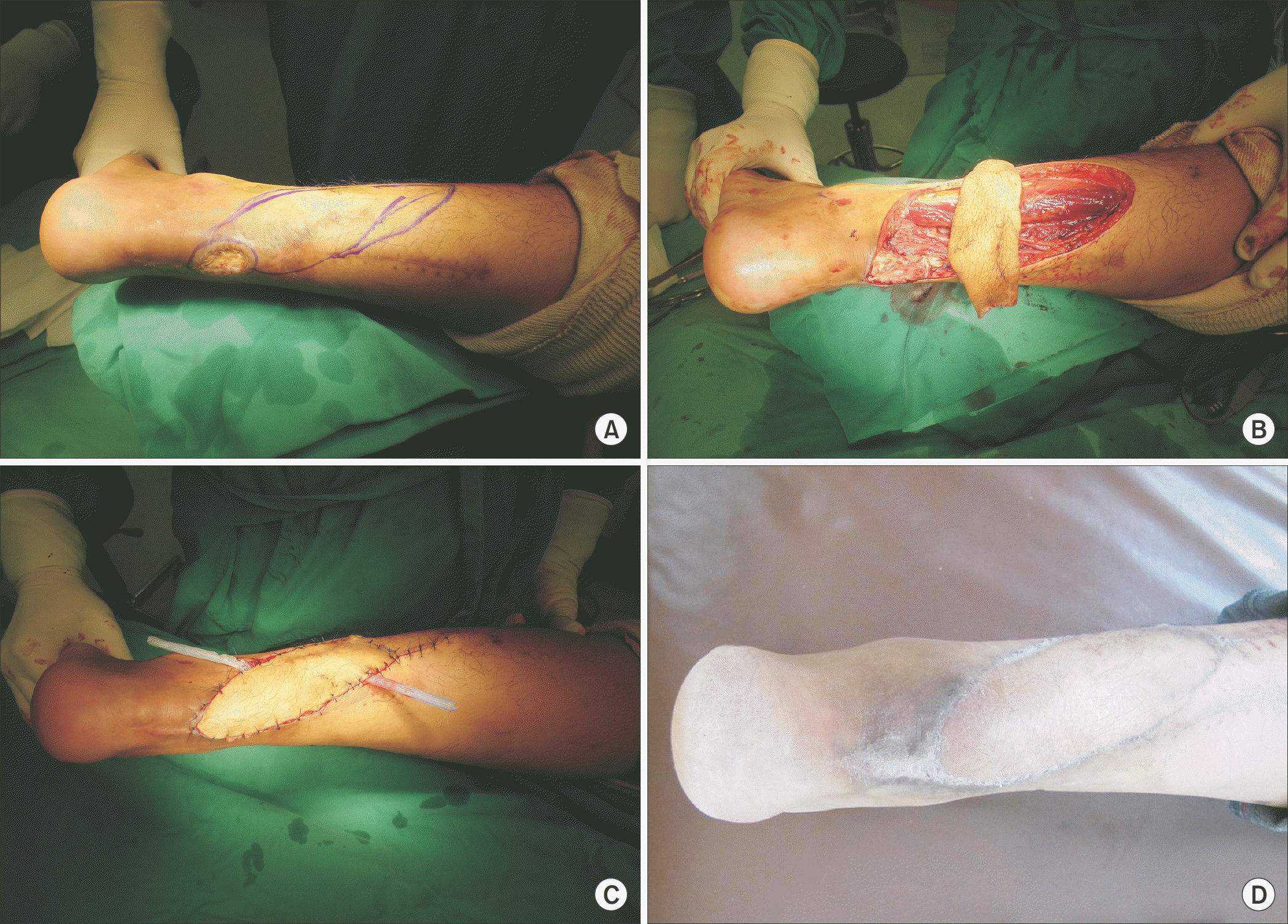
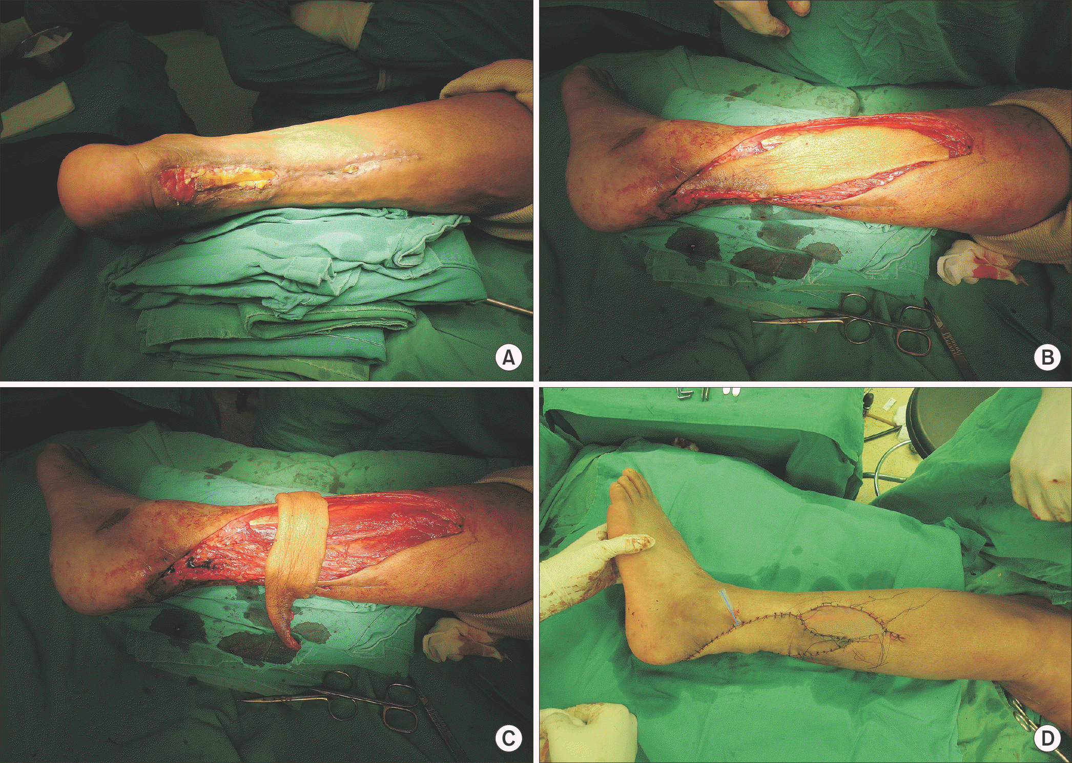
Figure 3.
These pictures show soft tissue defect and exposure of Achilles tendon after removal of devitalized tissue. (A) This picture shows preoperative state. (B) The flap was designed. (C) The flap was rotated with the perforator as a pivot. (D) The flap was inset and proximal wound was covered by skin graft.
 XML Download