

 PDF
PDF ePub
ePub Citation
 Print
Print


INTRODUCTION
Although several causes of lateral medullary infarction (LMI), such as atherosclerosis, dissection and thromboembolism in the intracranial vertebral artery (ICVA) and the posterior inferior cerebellar artery (PICA) were reported, there have been few radiological studies for ICVA and PICA in LMI as yet (1, 2).
Digital subtraction angiography (DSA) is a gold standard for diagnosis of arterial lesion in ICVA and PICA together with computed tomography angiography (CTA) and magnetic resonance angiography (MRA). But DSA is an inadequate method for screening arterial lesions due to invasiveness and the possibility of iatrogenic stroke (3). Lumen and thickened wall of cerebral artery are well evaluated using CTA with radiation risk. It is good to show luminal stenosis in intracranial artery in MRA, but have limitation to distinguish several diseases, which was shown as only luminal stenosis on MRA.
Recently advanced MR imaging techniques are available for detection and evaluation of atherosclerotic plaque and vessel wall in the intracranial artery. Several reports showed usefulness of high-resolution MRI in evaluation of plaques on intracranial artery such as basilar artery or middle cerebral artery (4, 5, 6, 7). High-resolution contrast-enhanced MRI helps to show intracranial arterial wall which is the small size and comparatively deep location (8). In past studies, high-resolution contrast-enhanced three dimensional gradient-echo image which was used to evaluate intracranial lesion in routine clinical examinations at 3T was more reliable than MRA for detecting ICVA dissection (9).
Therefore, the objectives of our study were to determine whether high-resolution contrast-enhanced three dimensional imaging with spoiled gradient-recalled sequence (HR-CE 3D-SPGR) plays a meaningful role in the assessment of ICVA and PICA in LMI patients.
MATERIALS AND METHODS
Patients
Twenty-five patients with LMI (19 men and 6 women; mean age, 58.5 years; range, 35-95 years) were enrolled from August 2006 to December 2012. This retrospective study was approved by the institutional review board of our institute. All patients underwent HR-CE 3D-SPGR and contrast-enhanced magnetic resonance angiography (CE-MRA) using a 3T MRI (GE Medical Systems) with variable time interval for imaging after symptom onset (mean time interval, 119.2 hours; range, 5-720 hours) and, diffusion weighted images (DWI) were used together with their clinical symptoms in order to confirm LMI. Sixteen patients had a previous history of hypertension. Nine patients had diabetes mellitus and seven had hyperlipidemia. Six patients had old cerebrovascular infarction. One patient had atrial fibrillation. Twelve patients had history of smoking. Among 25 patients, six patients showed vertigo and one patient showed loss of consciousness. Rest of them showed sensory change or motor weakness.
MR Imaging Protocol and Interpretation
Imaging was performed on a 3T MRI scanner (Signa Excite and Discovery MR750, GE Medical Systems, Milwaukee, Wisconsin, USA) with a standard eight-channel head coil.
CE MRA [TR/TE, 4.9/1.2; ST 1.4 mm; FOV 30 × 30 cm; matrix of 256 × 192, contrast material of 1.0 mmol of Gadobutrol (Gadovist; Bayer Schering Pharma, Berlin, Germany) per kilogram of body weight] were taken in all patients. In sequence, HR-CE 3D-SPGR [repetition time/echo time (TR/TE) msec of 6.9/1.7; flip angle of 20°; section thickness (ST) of 1 mm; field of view (FOV) of 20 × 20 cm; matrix of 256 × 256] was also obtained. Each HR-CE 3D-SPGR was transferred to the workstation software (Aquarius iNtuition Edition version 4.4.6.85.2800; TeraRecon, Inc, San Mateo, Calif). Transverse images perpendicular to vascular course of ICVA or PICA were obtained using this software for further evaluation.
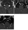
Arterial lesions in ICVA and PICA of LMI patients were evaluated with HR-CE 3D-SPGR and CE-MRA. Arterial lesions in ICVA on HR-CE 3D-SPGR were subcategorized as follows: 1. atheroma, 2. dissection, 3. thromboembolism, 4. no arterial lesion (Fig. 1). Atheroma on HR-CE 3D-SPGR was defined as thickness of wall between lumen showing high signal intensity (SI) and enhanced outer wall with additional consideration of simultaneous atheroma in intracranial vessels. Dissection on HR-CE 3D-SPGR was defined as double lumen containing true lumen and false lumen (10) with relatively intact other intracranial vessels. Thromboembolism in ICVA defined as very low SI arterial lesions with outer vessel wall, which meet neither atheroma nor dissection criteria. Arterial lesions in ICVA on CE-MRA were divided into subgroups as follows: 1. Stenosis, 2. Occlusion, 3. No arterial lesion. The presence or absence of arterial lesions in PICA was obtained on both HR-CE 3D-SPGR and CE-MRA.
It was divided into two groups of arterial lesion in ICVA relative to PICA involvement. Each group was compared about its demographics, the area of LMI, and cerebellar involvement. The area of LMI and lateral medulla were measured by manual tracing method in workstation. The extent of LMI was obtained from calculation of the area of LMI (ALMI) and lateral medulla (ALM) at the largest infarction level as follows: the area of LMI (%) = (ALMI/ALM) × 100.
These images were reviewed and analyzed independently blinded to clinical information and other brain MR images by two radiologists (Y.Y. and S.J.A.) with 3 and 6 years' experience of radiology. In a disagreement of opinion, two radiologists reached consensus with discussion.
Statistical Analysis
Mann-Whitney U test was calculated in continuous variables for comparison between two groups. Fischer exact test was used for comparison of categorical variables in each group. All statistical analyses were performed with SPSS (version 19.0, IBM, Chicago, Illinois, USA). P-value was defined as significant at less than 0.05.
RESULTS
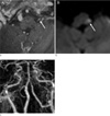
Two readers detected symptomatic side arterial lesions in ICVA or PICA in 22 patients with LMI on HR-CE 3D-SPGR. But CE-MRA showed arterial lesions in only 15 patients. Three cases were commonly not identified as arterial lesions in ICVA or PICA on two images by both two radiologists. Arterial lesions in only ICVA were detected in 10 LMI patients on HR-CE 3D-SPGR. CE-MRA also detected nine of ten only ICVA arterial lesions. One arterial lesion was missed on CE-MRA due to minimal atheroma in ICVA (Table 1). But PICA arterial lesions on CE-MRA were not visualized in 12 cases which were shown on HR-CE 3D-SPGR image (Fig. 2).
Among arterial lesions in ICVA on HR-CE 3D-SPGR image, there were 8 cases atheroma, 8 cases dissection and 2 cases thromboembolism (Table 2). Seven cases showed hypoplastic vertebral artery which was defined as less than 2 mm diameter in several literatures.
Group of ICVA arterial lesions with PICA involvement tended to simultaneously have cerebellar infarction with LMI (p = 0.069). The area of LMI between groups of ICVA arterial lesions with or without PICA involvement statistically showed no difference. Other factors were not different statistically (Table 3).
DISCUSSION
In LMI patients, we found that all arterial lesions in PICA which were shown on HR-CE 3D-SPGR image were not detected on CE-MRA. Concurrent cerebellar involvement was more shown in LMI patients with both ICVA and PICA arterial lesions than those with only ICVA arterial lesions.
There were several reports about causes of LMI with arterial lesions in ICVA or PICA (2, 11, 12). Fischer et al reported that pathologic finding of vessel involvements were 14.3% PICA disease, 38.1% VA disease and 26.2% both arteries diseases (13). Our results of 10 arterial lesions (40%) in only ICVA, 8 arterial lesions (32%) in both ICVA and PICA and 4 arterial lesions (16%) in only PICA were similar with that report. Our results showed 8 cases atheroma, 8 cases dissection, 2 cases thromboembolism on HR-CE 3D-SPGR, and 4 cases PICA lesions.
There were reports about non-invasive vessel imaging using CE-MRA which is able to depict lumen of intracranial artery such as ICVA (14, 15). However CE-MRA has technical limitation of the possibility in distinguishing of pathology in ICVA. In some cases, diffuse atherosclerosis or dissection in ICVA showed just luminal stenosis on CE-MRA. In one case, CE-MRA showed normal ICVA despite of presence of mild atherosclerosis. There was probably arterial remodeling that luminal narrowing did not occur until more than 50% atherosclerotic lesion in vessel wall existed (16, 17). There was a report that 3D-SPGR with T1W image was good to evaluate vertebral artery lesion such as dissection (10). To supplement this limitation, HR-CE 3D-SPGR was used for evaluation of arterial lesions in ICVA. In addition, our findings showed that HR-CE 3D-SPGR detected arterial lesions in PICA in 12 cases which were not shown on CE-MRA. HR-CE 3D-SPGR also helped to find arterial lesions in PICA which was small diameter using better spatial resolution than CE-MRA (18).
In our results cerebellar infarction was frequently shown in LMI patients with arterial lesion in ICVA with PICA involvement. Lateral medulla is largely supplied by distal vertebral artery and PICA (19). The PICA also supplies blood to the inferior cerebellum and pons (20). Some literatures which reported that PICA territory infarction was related with significant vertebral artery stenosis or occlusion supported our results (21, 22).
There were several limitations for this study. First, study with small population had limitation to make generalizations with our results. Although LMI was a rare disease, we believe that it was not small population to study LMI in light of prevalence (23). Second, absence of pathological confirmation for arterial lesion in ICVA or PICA caused incorrect results. But recent other studies showed possibility of evaluating intracranial small arteries such as basilar and middle cerebral artery using HR-MRI. These reports supported our study of evaluating arterial lesion in ICVA or PICA using HR-CE 3D-SPGR to enable detect small arterial lesion (4, 5, 6, 24). Third, it is difficult to distinguish hypoplastic vertebral artery from the pathology. There were some reports that these factors play an additional role in ischemic stroke (25, 26). Without stenosis, these factors were considered as risk factor for ischemia (12). In conclusion, HR-CE 3D-SPGR can help evaluate arterial lesions in ICVA and PICA for LMI patients.


 XML Download