

 PDF
PDF ePub
ePub Citation
 Print
Print


Abstract
Purpose
We compared non-invasive imaging studies of CTA, TOF-MRA and CE-MRA to evaluate detecting internal carotid artery stenosis and occlusion.
Materials and Methods
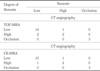
We enrolled 20 patients with clinically suspected internal carotid artery stenosis and occlusion, or asymptomatic patient with more than 50% stenosis suspected on color Doppler ultrasonography for regular check-up. Prospectively, CTA, TOF-MRA and CE-MRA were performed, and sensitivity, specificity, positive predictive value, negative predictive value and accuracy of TOF-MRA and CE-MRA using CTA as a reference standard for detecting more than 50% stenosis were evaluated, and correlations of measured percent stenosis between 3 imaging studies were also evaluated.
Results
No significant difference was found between 3 imaging studies in measuring stenosis(p>0.05). Correlation coefficient was 0.932 between CTA and TOF-MRA, and 0.971 between CTA and CE-MRA. TOF-MRA had 83.3% sensitivity, 87.5% specificity, 71.4% positive predictive value, 93.3% negative predictive value and 86.4% accuracy for detecting more than 50% stenosis and occlusion. CE-MRA had 83.3% sensitivity, 93.8% specificity, 83.3% positive predictive value, 93.8% negative predictive value and 90.9% accuracy.
Figures and Tables
Fig. 1
A 73-year-old male patient with headache and dizziness.
(a) TOF-MRA (b) CE-MRA (c) CTA, These images show a low grade stenosis in right ICA.
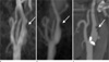
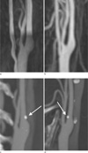
Fig. 2
A 64-year-old male non-symptomatic patient.
(a) TOF-MRA shows no stenotic lesion or plaque.
(b) CE-MRA shows no stenotic lesion or plaque.
(c, d) CTA(C: coronal, D: sagittal) shows a small calcified plaque in the left proximal ICA with mild degree stenosis.
References
1. Statistics Korea. 2009.
2. Randoux B, Marro B, Koskas F, Duyme M, Sahel M, Zouaoui A, et al. Carotid artery stenosis: prospective comparison of CT, three-dimensional gadolinium-enhanced MR, and conventional angiography. Radiology. 2001. 220:179–185.
3. Mühlenbruch G, Das M, Mommertz G, Schaaf M, Langer S, Mahnken AH, et al. Comparison of dual-source CT angiography and MR angiography in preoperative evaluation of intra- and extracranial vessels: a pilot study. Eur Radiol. 2010. 20:469–476.
4. Babiarz LS, Romero JM, Murphy EK, Brobeck B, Schaefer PW, Gonzlez RG, et al. Contrast-enhanced MR angiography is not more accurate than unenhanced 2D time-of-flight MR angiography for determining > or = 70% internal carotid artery stenosis. AJNR Am J Neuroradiol. 2009. 30:761–768.
5. Alvarez Linera J, Benito-Len J, Escribano J, Campollo J, Gesto R. Prospective evaluation of carotid artery stenosis: elliptic centric contrast-enhanced MR angiography and spiral CT angiography compared with digital subtraction angiography. AJNR Am J Neuroradiol. 2003. 24:1012–1019.
6. Sadikin C, Teng MM, Chen T, Luo C, Chang F, Lirng J, et al. The current role of 1.5T non-contrast 3D time-of-flight magnetic resonance angiography to detect intracranial steno-occlusive disease. Journal of the Formosan Medical Association. 2007. 106:691–699.
7. Lell M, Fellner C, Baum U, Hothorn T, Steiner R, Lang W, et al. Evaluation of carotid artery stenosis with multisection CT and MR imaging: influence of imaging modality and postprocessing. AJNR Am J Neuroradiol. 2007. 28:104–110.
8. Villablanca JP, Nael K, Habibi R, Nael A, Laub G, Finn JP. 3 T contrast-enhanced magnetic resonance angiography for evaluation of the intracranial arteries: comparison with time-of-flight magnetic resonance angiography and multislice computed tomography angiography. Invest Radiol. 2006. 41:799–805.
9. Jang NK, Seo JJ, Chung TW, Jeong GW, Kim JK, Kang HK, Cho KH. The Usefulness of Enhanced 3D-TOF MR Angiography in the Patients with Cerebral Infarction: Comparison with Conventional Angiography. J Korean Radiol Soc. 2000. 42:575–583.
10. Atkinson D, Brant Zawadzki M, Gillan G, Purdy D, Laub G. Improved MR angiography: magnetization transfer suppression with variable flip angle excitation and increased resolution. Radiology. 1994. 190:890–894.
11. Townsend TC, Saloner D, Pan XM, Rapp JH. Contrast material-enhanced MRA overestimates severity of carotid stenosis, compared with 3D time-of-flight MRA. J Vasc Surg. 2003. 38:36–40.
12. DeMarco JK, Huston J, Bernstein MA. Evaluation of classic 2D time-of-flight MR angiography in the depiction of severe carotid stenosis. AJR Am J Roentgenol. 2004. 183:787–793.
13. Rasanen HT, Manninen HI, Vanninen RL, Vainio P, Berg M, Saari T. Mild carotid artery atherosclerosis: assessment by 3-dimensional time-of-flight magnetic resonance angiography, with reference to intravascular ultrasound imaging and contrast angiography. Stroke. 1999. 30:827–833.
14. Evans AJ, Richardson DB, Tien R, MacFall JR, Hedlund LW, Heinz ER, et al. Poststenotic signal loss in MR angiography: effects of echo time, flow compensation, and fractional echo. AJNR Am J Neuroradiol. 1993. 14:721–729.
15. Sardanelli F, Zandrino F, Parodi RC, De Caro G. MR angiography of internal carotid arteries: breath-hold Gd-enhanced 3D fast imaging with steady-state precession versus unenhanced 2D and 3D time-of-flight techniques. J Comput Assist Tomogr. 1999. 23:208–215.
16. Kramer H, Runge VM, Morelli JN, Williams KD, Naul LG, Nikolaou K, et al. Magnetic resonance angiography of the carotid arteries: comparison of unenhanced and contrast enhanced techniques. Eur Radiol. 2011.
17. Morcos SK. Nephrogenic systemic fibrosis following the administration of extracellular gadolinium based contrast agents: is the stability of the contrast agent molecule an important factor in the pathogenesis of this condition? Br J Radiol. 2007. 80:73–76.
18. Grobner T. Gadolinium--a specific trigger for the development of nephrogenic fibrosing dermopathy and nephrogenic systemic fibrosis? Nephrol Dial Transplant. 2006. 21:1104–1108.
19. Port M, Ide J, Medina C, Robic C, Sabatou M, Corot C. Efficiency, thermodynamic and kinetic stability of marketed gadolinium chelates and their possible clinical consequences: a critical review. Biometals. 2008. 21:469–490.
20. Morcos SK. Extracellular gadolinium contrast agents: differences in stability. Eur J Radiol. 2008. 66:175–179.
 XML Download