

 PDF
PDF ePub
ePub Citation
 Print
Print


INTRODUCTION
The obstructed hemivagina and ipsilateral renal anomaly (OHVIRA) syndrome is a rare congenital anomaly of the Müllerian (paramesonephric) and Wolffian (mesonephric) ducts. It was initially reported in 1922 by Purslow and also referred to as the Herlyn-Werner-Wunderlich syndrome. It typically occurs concurrently with uterine didelphys and ipsilateral renal agenesis (12345). Ipsilateral renal agenesis can be accompanied with other urologic anomalies simultaneously. A blind ectopic ureter combined with ipsilateral agenesis and Müllerian duct anomalies is extremely rare (67). To our knowledge, a blind ectopic ureter associated with OHVIRA syndrome has not been reported in English literature. The diagnosis of ureteral anomalies can be challenging due to their rare incidence and cystic features that can be mistaken for an adnexal lesion in female patients. Regarding this matter, MRI is considerably helpful not only for diagnosing Müllerian duct anomalies, but also for detecting and characterizing associated non-uterine anomalies (12345). In this case report, we present an unusual case of a 13-year-old girl with OHVIRA syndrome and a blind ectopic megaureter, which was successfully diagnosed using MRI.
CASE REPORT
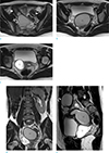
A 13-year-old female was presented to our emergency department with complaints of acute lower abdominal pain. Her menarche occurred at the age of 12, and she had regular menstrual cycles, once every 30 days for 4 to 5 days. She reported mild dysmenorrhea, which was manageable without medications. Physical examination revealed severe tenderness over the right lower quadrant of the abdomen. The patient was afebrile with normal vital signs. Laboratory findings, including complete blood count and urine analysis, were normal, and a pregnancy test was negative. She underwent computed tomography (CT) with clinical suspicion of early appendicitis. Abdominal and pelvic CT revealed two uterine horns, which were widely separated, and a large high-attenuated cystic mass at the lower aspect of both uterine horns. The right uterus was distended with slightly high-attenuated fluid and was clearly connected to the cystic mass, consistent with a hematometrocolpos. The communication between the left uterus and the hematocolpos was not clearly visualized, which suggested the possibility of a bicornuate uterus or uterine didelphys. A dilated tubular structure filled with slightly high-attenuated fluid and in close proximity to the right uterine horn, was also observed in the right side of the pelvis, consistent with a hematosalpinx. A well-defined and thin walled, unilocular cystic mass was seen posterior to the right aspect of the urinary bladder, and it was suspected to be a dilated blind ectopic ureter or cystic ovarian mass. The right kidney was not visualized, and the left kidney was relatively hypertrophied. Magnetic resonance imaging (MRI) was performed for further evaluation of a uterine abnormality and the cystic mass posterior to the urinary bladder. MRI showed two completely separate uteruses with normal zonal anatomy (Fig. 1a), two separate cervices, and a large hematocolpos connected to the right uterus, corresponding to the obstructed right hemivagina (Fig. 1b-e). Right-sided hematometra and hemotosalpinx (Fig. 1e) were also seen. The left uterus had its own opening into the left hemivagina (Fig. 1b-d). Both ovaries appeared normal, and the right kidney was absent (Fig. 1d). The cystic mass, which was located at the right lower pelvic cavity posterior to the bladder, had no communication with the right uterus or the hemivagina and was filled with fluid, which was hyperintense on the T2-weighted image (Fig. 1c-e) and hypointense on the T1-weigted image. Although an opening tract between the cystic mass and the bladder was not clearly visible, the lesion was most likely a dilated blind ectopic ureter. On the basis of the imaging findings of uterine didelphys, unilateral obstructed hemivagina with resultant hematometrocolpos and hematosalpinx, and unilateral renal agenesis, the case was diagnosed as OHVIRA syndrome.
Vaginal septotomy and drainage of hematometrocolpos and hematosalpinx were performed, and the patient was discharged without any immediate complications. Three months later, the patient complained of progressive lower abdominal pain. Hysteroscopy revealed stenosis of the vaginal septum, and drainage of hematocolpos was performed with dilatation of vaginal septotomy. However, the patient later underwent right hysterectomy because the symptom was not relieved. The cystic mass, which was located posterior to the urinary bladder, was also excised, and it was diagnosed as a blind megaureter. At the 18-month follow-up, the patient had no complications and had a normal menstrual flow.
DISCUSSION
The pathogenesis and etiology of OHVIRA syndrome is still unclear. It has been considered to be the result of a developmental anomaly of the Müllerian and Wolffian ducts (1234). Two pairs of Müllerian ducts fuse craniocaudally to form the uterus, cervix, and upper two-thirds of the vagina. Also, fused Müllerian ducts perforate into the urogenital sinus, which gives rise to the lower third of the vagina. Wolffian ducts not only develop into the ureter and the kidneys, but also induce adequate Müllerian duct fusion. Maldevelopment of the Wolffian duct causes failure of ureter and kidney differentiation, as well as a lateral displacement of the ipsilateral Müllerian duct. The displaced Müllerian duct cannot fuse with the contralateral duct, causing uterine didelphys, and the resultant failure to contact the urogenital sinus forms a blind sac, leading to an obstructed hemivagina (1234).
In our case, the blind ectopic megaureter was seen in combination with ipsilateral renal agenesis. This anomaly may also be explained by maldevelopment of the Wolffian duct, which gives rise to the ureteric bud (67).
While typical presentation of OHVIRA syndrome includes ipsilateral renal agenesis, a variety of associated urologic anomalies have been reported, including multicystic dysplastic kidneys, ectopic ureters, and duplication of kidneys and ureters (45). Renal anomalies are almost always located on the same side as the obstructed hemivagina, and the right side is affected twice as often as the left side (123).
The OHVIRA syndrome generally occurs shortly after menarche, and patients usually present with pelvic pain or dysmenorrhea, with or without palpable mass due to hematocolpos or hematometra (1). In incomplete obstruction, clinical presentation can be delayed as there remains one patent hemivagina, allowing for menstrual blood to exit, while the other side is obstructed (12). As the incidence of complications including endometriosis, menstrual disorders, infertility, and obstetric complications increases with time (123), accurate and early diagnosis is essential.
Ultrasound and MRI are the most commonly used imaging modalities in the diagnosis of Müllerian duct anomalies. Ultrasound is frequently used as an initial examination in obstetric and gynecologic evaluations, as it is noninvasive, free of radiation, and widely available. However, it cannot identify the types of Müllerian duct anomalies (348). On the other hand, MRI is considered the ideal imaging modality for noninvasive evaluation of female pelvic anatomy because it provides more detailed information regarding uterine morphology, the continuity with each vaginal lumen, the character of the vaginal septum, and the nature of fluid contents. It is also more useful, when compared to other imaging modalities, in the detection of associated conditions such as endometriosis, pelvic inflammation and adhesions, and coexisting urologic anomalies (123457).
Resection of the obstructed vaginal septum is the treatment of choice for symptom relief and preservation of reproductive capability (159). Hemihysterectomy with or without salphingo-oophorectomy should be avoided as the reported incidence of pregnancy in both horns is almost equal. Hemihysterectomy is only used in selected cases for which resection of the vaginal septum is not enough to relieve the hematometrocolpos or recurrent vaginal stenosis develops, as in our case (19).
In conclusion, MRI is the most powerful tool in establishing the diagnosis of Müllerian duct anomalies and associated conditions, when compared to other imaging modalities. MRI must be performed in young females suspected to have OHVIRA syndrome in order to make an accurate diagnosis and to optimize the appropriate treatment.
 XML Download