

 PDF
PDF ePub
ePub Citation
 Print
Print


Abstract
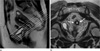
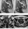
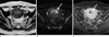
Although ultrasonography is the most commonly used first-line imaging modality of the female pelvis because of diagnostic accuracy, low cost and safety, MRI is the best imaging modality of choice for the evaluation of the female pelvis. The indication of female pelvis MRI is diverse and includes the evaluation of Müllerian duct anomaly, differential diagnosis and characterization of uterine and ovarian tumors, and staging of malignant uterine and ovarian tumors. Understanding of MR protocols according to the specific gynecologic pathology allows accurate diagnosis and proper patient management.
Figures and Tables
References
1. Brown MA, Mattrey RF, Stamato S, Sirlin CB. MRI of the female pelvis using vaginal gel. AJR Am J Roentgenol. 2005. 185:1221–1227.
2. Taupitz M, Rogalla P. Hamm B, Forstner R, editors. MR and CT techniques. MRI and CT of the female pelvis. 2007. Heidelberg: Springer-Verlag;25–36.
3. Baudouin CJ, Soutter WP, Gilderdale DJ, et al. Magnetic resonance imaging of the uterine cervix using an intravaginal coil. Magn Reson Med. 1992. 24:196–203.
4. Rofsky NM, Lee VS, Laub G, et al. Abdominal MR imaging with a volumetric interpolated breath-hold examination. Radiology. 1999. 212:876–884.
5. Ascher SM, O'Malley J, Semelka RC, Patt RH, Rajan S, Thomasson D. T2-weighted MRI of the uterus: fast spin echo vs breat-hold fast spin echo. J Magn Reson Imaging. 1999. 9:384–390.
6. Masui T, Katayama M, Kobayashi S, Sakahara H, Ito T, Nozaki A. T2-weighted MRI of the female pelvis: comparison of breath-hold fast-recovery fast spin-echo and nonbreath-hold fast spin-echo sequences. J Magn Reson Imaging. 2001. 13:930–937.
7. Lichy MP, Wietek BM, Mugler JP 3rd, et al. Magnetic resonance imaging of the body trunk using a single-slab, 3-dimensional, T2-weighted turbo-spin-echo sequence with high sampling efficiency (SPACE) for high spatial resolution imaging: initial clinical experiences. Invest Radiol. 2005. 40:754–760.
8. Bellin MF. MR contrast agents, the old and the new. Eur J Radiol. 2006. 60:314–323.
9. Prince MR, Zhang HL, Prowda JC, Grossman ME, Silvers DN. Nephrogenic systemic fibrosis and its impact on abdominal imaging. Radiographics. 2009. 29:1565–1574.
10. Perez-Rodriguez J, Lai S, Ehst BD, Fine DM, Bluemke DA. Nephrogenic systemic fibrosis: incidence, associations, and effect of risk factor assessment--report of 33 cases. Radiology. 2009. 250:371–377.
11. Troiano RN, McCarthy SM. Müllerian duct anomalies: imaging and clinical issues. Radiology. 2004. 233:19–34.
12. Kinkel K, Kaji Y, Yu KK, et al. Radiologic staging in patients with endometrial cancer: a meta-analysis. Radiology. 1999. 212:711–718.
13. Yamashita Y, Harada M, Sawada T, Takahashi M, Miyazaki K, Okamura H. Normal uterus and FIGO stage I endometrial carcinoma: dynamic gadolinium-enhanced MR imaging. Radiology. 1993. 186:495–501.
14. Hricak H, Finck S, Honda G, Goranson H. MR imaging in the evaluation of benign uterine masses: value of gadopentetate dimeglumine-enhanced T1-weighted images. AJR Am J Roentgenol. 1992. 158:1043–1050.
15. Hricak H, Chen M, Coakley FV, et al. Complex adnexal masses: detection and characterization with MRI--multivariate analysis. Radiology. 2000. 214:39–46.
16. Wu TT, Coakely FV, Qayyum A, et al. Magnetic resonance imaging of ovarian cancer arising in endometriomas. J Comput Assist Tomogr. 2004. 28:836–838.
17. Kurihara Y, Yakushiji YK, Tani I, Nakajima Y, Van Cauteren M. Coil sensitivity encoding in MR imaging: advantages and disadvantages in clinical practice. AJR Am J Roentgenol. 2002. 178:1087–1091.
18. Koyama T, Umeoka S, Kataoka M, et al. Utility of diffusionweighted MR imaging in evaluation of esophageal cancers. 2005. In : Proceedings of the 13th Annual Meeting of ISMRM; Miami Beach, FL, USA. 2820.
19. Namimoto T, Awai K, Nakaura T, Yanaga Y, Hirai T, Yamashita Y. Role of diffusion-weighted imaging in the diagnosis of gynecological disease. Eur Radiol. 2009. 19:745–760.
20. Whittaker CS, Coady A, Culver L, Rustin G, Padwick M, Padhani AR. Diffusion-weighted MR imaging of female pelvis tumors: a pictorial review. Radiographics. 2009. 29:759–774.
21. Rechichi G, Galimberti S, Signoreli M, Perego P, Valsecchi MG, Sironi S. Myometrial invasion in endometrial cancer: diagnostic performance of diffusion-weighted MR imaging at 1.5-T. Eur Radiol. 2010. 20:754–762.
22. Fujii S, Matsuse E, Kanasaki Y, et al. Detection of peritoneal dissemination in gynecological malignancy: evaluation by diffusion-weighted MR imaging. Eur Radiol. 2008. 18:18–23.
23. Thomassin-Naggara I, Darai E, Cuenod CA, et al. Contribution of diffusion-weighted MR imaging for predicting benignity of complex adnexal masses. Eur Radiol. 2009. 19:1544–1552.
24. Lim RP, Lee VS, Bennett GL, et al. Imaging of the female pelvis at 3.0T. Top Magn Reson Imaging. 2006. 17:427–443.
25. Cornfeld D, Weinreb J. Simple changes to 1.5-T MRI abdomen and pelvis protocols to optimize results at 3T. AJR Am J Roentgenol. 2008. 190:W140–W150.
26. Brown MA, Martin DR, Semelka RC. Future directions in MR imaging o the female pelvis. Magn Reson Imaging Clin N Am. 2006. 14:431–437.
 XML Download