

 PDF
PDF Citation
 Print
Print


INTRODUCTION
Traumatic brain injury (TBI) is a major cause of mortality and disability. TBI is classified as mild, moderate, and severe based on the severity and 70%–90% cases of TBI are classified as mild TBI [123]. The incidence of hospital-treated patients with mild TBI is approximately 100–300/100,000 population, however, the true population-based rate is probably above 600/100,000 when including mild TBI not treated in hospitals [4].
Despite having different definitions, cerebral concussion and mild TBI have been used interchangeably, therefore the terminology regarding head trauma has caused confusion for patients and doctors [5]. Since the development of diffusion tensor imaging (DTI), many studies have demonstrated traumatic axonal injury (TAI) lesions in patients with concussion or mild TBI [67891011121314]. TAI is a more severe TBI subtype than concussion or mild TBI and use of TAI is rare in Korea, thus there has been significant debate and confusion in the clinical field of Korea (Table 1). We think that reviewing the history of terminology of ‘TAI’ with concussion and mild TBI would be helpful to clarifying this debate and confusion in Korea.
Table 1
Traumatic brain injury subtypes

In this review article, we reviewed the history of TAI in patients with concussion and mild TBI.
DEFINITION OF CEREBRAL CONCUSSION, MILD TBI, AND TRAUMATIC AXONAL INJURY
Cerebral concussion is defined as a transient, temporary, neurological dysfunction resulting from application of force to the brain: in detail, cerebral concussion is an acute trauma-induced change of mental function generally lasting less than 24 hours and usually recovering within 2–3 weeks [515]. Concussion is not usually associated with visible lesions that can be detected by conventional CT or MRI [5161718].
In 1993, the American Congress of Rehabilitation Medicine defined mild TBI as a traumatically induced physiological disruption of brain function resulting from the head being struck or striking an object or an acceleration and deceleration movement of the brain, as manifested by at least one of the following: any period of loss of consciousness up to 30 minutes; post-traumatic amnesia not exceeding 24 hours and a Glasgow Coma Scale score of 13–15 [16192021]. In 1995, Alexander added two more conditions for diagnosis of mild TBI: the patient has no focal signs and conventional neuroimaging studies are negative [16].
TAI is a more severe subtype of TBI than concussion or mild TBI (Table 1) [22]. Neural axons in the white matter appear to be particularly vulnerable to diffuse head injury due to the mechanical loading of the brain during TBI [2324]. TAI is defined as tearing of axons due to indirect shearing forces during acceleration, deceleration and rotation of the brain, or direct head trauma [6789101112131425262728].
There have been opposing opinions regarding the use of the terms “concussion” and “mild TBI.” Sharp and Jenkins [18] insisted that mild TBI is not always a benign condition as its name implies and patients sometimes fail to recover. They proposed that the term “concussion” should be avoided because it has no clear definition and no pathological meaning, therefore, at worst it encourages an apathetic diagnostic approach [18]. McMahon et al. [29] insisted that the term “mild TBI” is a misnomer for patients with severe post-concussion syndrome. Rapp and Curley [30] reported that mild TBI is a category mistake because of the heterogeneity of the clinical population and clinical presentations, the absence of a unitary etiology of post injury deficits, and the complex idiosyncratic time course of the appearance of these deficits in mild TBI.
PROBLEMS IN DIAGNOSIS OF CEREBRAL CONCUSSION AND MILD TBI
Cerebral concussion is a transient disorder of brain function without long-term sequelae [18]. Therefore, patients with concussion should make a complete recovery with no sequelae. However, a significant proportion of patients with concussion showed sequelae with a reported incidence of approximately 15% at one year following concussion [31]. These patients were grouped as post-concussion syndrome, which has been regarded as a psychological problem [3132]. By contrast, in a recent large multicenter study conducted in America and England, McMahon et al. [29] reported that 82% of patients reported at least one post-concussion syndrome and 22.4% of patients were still below functional state at 12 months after onset. As a result, the opinion that post-concussion syndrome is not a psychological problem but a physical problem, especially TAI, has been suggested [93233]. Messé et al. [33], who detected TAI lesions using DTI in patients with poor outcome following mild TBI, suggested that post-concussion syndrome might be a consequence of TAI. Other researchers have also suggested a possible association between post-concussion syndrome and TAI [932].
It is well-known that conventional CT and MRI are not sensitive to detection of TAI in patients with concussion or mild TBI [934]. Most problems in diagnosis of TAI in patients with concussion or mild TBI have originated from this insensitivity of conventional CT and MRI. By contrast, since development of DTI, many studies have detected TAI lesions in patients with mild TBI who showed normal conventional CT or MRI [67891011121314]. Based on these results, normal conventional CT or MRI findings in patients with concussion or mild TBI is not an indication that the patient’s brain is in a normal state without TAI lesions, therefore, conventional CT and MRI should not be mainly used for diagnosis of TAI in patients with concussion or mild TBI [934].
HISTORY OF TRAUMATIC AXONAL INJURY IN CONCUSSION AND MILD TBI
TAI has been used for several decades as a subtype of TBI [24]. However, in Korea, it has rarely been used in the clinical field while diffuse axonal injury (DAI) and concussion have been widely used. Since the middle of the last century, several studies have demonstrated trauma related axonal injury in the human brain using autopsy [35]. In 1982, Adams et al. [36], who introduced the term “DAI” by characterizing the extent and distribution of axonal injury in patients with TBI, defined DAI as the presence of microscopic axonal injury in the white matter of the cerebral hemisphere, corpus callosum, and brainstem caused by mechanical forces following head trauma [3738]. Many subsequent studies have reported DAI in patients with moderate or severe TBI. Efforts have been made to use terms including “TAI” or “diffuse TAI” to correct the false term “diffuse” (actual distribution of axonal injury lesion is not diffuse but multifocal) and underline the etiology of the axonal injury in trauma instead of DAI [2324]. On the other hand, since the 1980’s, many researchers, including Povlishock, have used the term “TAI” in their histopathological studies [23252639]. Patients with the traditional definition of DAI are in profound coma from the onset of injury and usually have a poor outcome [3640]. Because patients have more restricted patterns of axonal injury than that seen in the classic DAI with the development of neuroimaging techniques, the term “TAI” has been used for these more limited injuries: in practice, TAI has been used in milder cases than DAI [40]. The current tendency is to use one term “TAI” including DAI and the unification of terms “TAI” and “DAI” is necessary to make clear confusion and debate for these terms.
Regarding the evidence of TAI lesions in patients with concussion or mild TBI, since the 1960’s, several studies have reported on TAI in patients with concussion who showed no radiological evidence of brain injury by autopsy [414243]. One famous study was published by Lancet in 1994: Blumbergs et al. [42] reported on detection of axonal injury by autopsy in the brain of 5 patients with concussion, who died of other causes. However, conventional CT and MRI are not sensitive to detection of axonal injury in concussion or mild TBI, therefore, previously, diagnosis of TAI in live patients with concussion or mild TBI could not be demonstrated.
With the development of DTI in the 1990’s, many studies have demonstrated the usefulness of DTI in detection of TAI and TAI lesions in live animals and in human brain with concussion or mild TBI [67891011121314]. In 2007, Mac Donald et al. [44], who demonstrated the usefulness of DTI for diagnosis of TAI in a mouse model of mild TBI which showed normal findings on conventional MRI, concluded that DTI is really sensitive for detection of TAI and conventional MRI is not as sensitive as DTI for axonal injury. Regarding the demonstration of TAI in live human patients with concussion or mild TBI, in 2002, Arfanakis et al. [6] reported on TAI lesions in patients with mild TBI using DTI for the first time. Subsequently, TAI has been demonstrated in patients with concussion or mild TBI in hundreds of studies [67891011121314].
DTI AND DTI TRACTOGRAPHY
DTI provides invaluable information about subcortical white matter not available with conventional CT and MRI, thus development of DTI in the 1990’s has led to a new era for examination of the subcortical white matter in the live human brain [94546]. DTI is a sensitive measure of axonal injury that is particularly important for evaluation of small and subtle brain alterations in patients with mild TBI [9]. DTI was first employed in examination of white matter pathology in various other brain pathologies, including multiple sclerosis, stroke, Alzheimer’s disease, and schizophrenia [9]. Regarding mild TBI, Arfanakis et al. [6], the first researchers to use DTI for examination of TAI in patients with mild TBI, demonstrated TAI lesions in the subcortical white matter in five patients with mild TBI; they concluded that DTI is a powerful technique for evaluation of axonal injury in mild TBI. Subsequently, many researchers including Inglese et al. [7], Niogi et al. [8], and so on have reported on TAI in patients with mild TBI [69]. Recently, in a review article on DTI and mild TBI, Shenton et al. [9] described DTI as the best imaging technique available for detection of subcortical white matter damage, therefore, DTI will likely become an important diagnostic tool for patients with mild TBI, particularly in cases where conventional CT and MRI are negative.
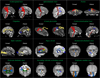
DTI tractography, which is reconstructed from DTI data, was developed for visualization and estimation of the neural tracts in the subcortical white matter of the brain [47]. The main advantage of DTI tractography is that the whole neural tract, instead of just a portion of the neural tract, can be evaluated [9]. Therefore, DTI tractography is a promising tool that can be used to find where damage occurs along the neural tracts [9]. As a result, DTI tractography can be used for both visualization and quantification of injury of the neural tracts in the subcortical white matter in a single patient and thus these methods are potentially important for diagnosis of TAI in patients with concussion or mild TBI [9]. Recently, TAI of various neural tracts in patients with mild TBI has been demonstrated in tens of studies: these neural tracts include the corticospinal tract, spinothalamic tract, fornix, cingulum, optic radiation, and so on (Fig. 1) [1011121314].
CONCLUSION
In this review article, we reviewed history of TAI in cerebral concussion and mild TBI. The introduction of DTI has enabled accurate diagnosis of TAI in patients with concussion or mild TBI. Therefore, rare use of TAI in Korea appeared to be related to slow development of DTI analysis techniques in Korea. Because TAI is a more severe TBI subtype than concussion and mild TBI, accurate diagnosis of TAI in patients with concussion or mild TBI is important for the following problems: false diagnosis of TAI with milder TBI subtype (concussion and mild TBI), delayed diagnosis of TAI, and loss of the critical period for recovery following TAI. Therefore, we think that use of DTI analysis technique for diagnosis of TAI should be facilitated in Korea.
 XML Download