

 PDF
PDF ePub
ePub Citation
 Print
Print


Abstract
Purpose
The goal of this study using the biomechanical test was to evaluate the mechanical stability of the bone-plate fixation system according to changes of the fracture gap sizes and widths.
Materials and Methods
For mechanical test, four types with different fracture models simulating the clinical situations were constructed depending on the gap size (FGS, mm) and the gap width (FGW, %) at the fracture site: 0 mm/0%, 1 mm/100%, 4 mm/100%, 4 mm/50%. For analyzing the effects of fracture gap on the biomechanical stability of the bone-plate fixation system, 4-point bending test was performed under all same conditions.
Results
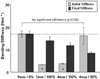
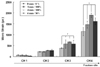
It was found that the fracture gap sizes of 1 and 4 mm decreased mechanical stiffness by about 50~60% or more. Furthermore, even without fracture gap size, 50% or more fracture gap width considerably decreased mechanical stiffness and suggested the possibility of plate damage through strain results.
Figures and Tables
Figure 1
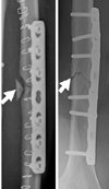
The LC-DCP bone fixation system and the epoxy pipe represented as a bone (A) and specimen configurations (B) for the fracture gap sizes & width (mm/%).
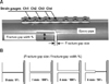
Figure 2
Mechanical experiment through 4-pont bending test (A→B→D→C) with LC-DCP bone- plate fixation system.
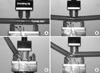
Figure 3
Determination of the initial and final bending stiffness (Nm/°) with the bending moment (Nm)-bending angle (°) curve.
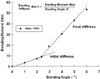
Figure 4
Comparison of bending moments according to the fracture gap sizes and widths on the LC-DCP fixation system.
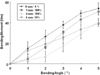
Figure 5
Comparison of the initial and final bending stiffness (Nm/°) according to the fracture gap sizes and widths of LC-DCP: *indicates significant difference compared with 0 mm/0% model in the initial stiffness.
References
1. Baumgaertel F, Buhl M, Rahn BA. Fracture healing in biological plate osteosynthesis. Injury. 1998. 29:Suppl 3. C3–C6.

2. Cordey J, Borgeaud M, Perren SM. Force transfer between the plate and the bone: relative importance of the bending stiffness of the screws and the friction between plate and bone. Injury. 2000. 31:Suppl 3. C21–C28.
3. Frigg R. Locking compression plate (LCP). An osteosynthesis plate based on the Dynamic Compression Plate and Point Contact Fixator (PC-Fix). Injury. 2001. 32:Suppl 2. 63–66.
4. Gardner MJ, Brophy RH, Campbell D, et al. The mechanical behavior of locking compression plates compared with dynamic compression plates in a cadaver radius model. J Orthop Trauma. 2005. 19:597–603.
5. Gautier E, Perren SM, Cordey J. Effect of plate position relative to bending direction on the rigidity of a plate ostosynthesis. A theoretical analysis. Injury. 2000. 31:Suppl 3. C14–C20.
6. Korner J, Diederichs G, Arzdorf M, et al. A biomechanical evaluation of methods of distal humerus fracture fixation using locking compression plate versus conventional reconstruction plates. J Orthop Trauma. 2004. 18:286–293.
7. Fulkerson E, Egol KA, Kubiak EN, Liporace F, Kummer FJ, Koval KJ. Fixation of diaphyseal fractures with segmental defects: a biomechanical comparison of locked and conventional plating techniques. J Trauma. 2006. 60:830–835.
8. Perren SM. Evolution of the internal fixation of long bone fracture. J Bone Joint Surg Br. 2002. 84:1093–1110.
9. Ramakrishna K, Shidhar I, Sivashanker S, Khong KS, Ghista DN. Design of fracture fixation plate for necessary and sufficient bone stress shielding. JSME International Journal Series C. 2004. 47:1086–1094.
10. Sommer C, Gautier E, Müller M, Helfet DL, Wagner M. First clinical results of the locking compression plate (LCP). Injury. 2003. 34:Suppl 2. B43–B54.
11. Stoffel K, Dieter U, Stachowiak G, Gächter A, Kuster MS. Biomechanical testing of the LCP - how can stability in locked internal fixators be controlled? Injury. 2003. 34:Suppl 2. B11–B19.
12. Stoffel K, Stachowiak G, Forster T, Gächter A, Kuster M. Oblique screws at the plate ends increase the fixation strength in synthetic bone test medium. J Orthop Trauma. 2004. 18:611–616.
 XML Download