

 PDF
PDF ePub
ePub Citation
 Print
Print


INTRODUCTION
Nitrogen-containing bisphosphonates (BPs), such as pamidronate, alendronate, and risedronate, can inhibit osteoclastic function and reduce osteoclast number by inducing apoptotic cell death in osteoclasts.[1234] Long-term BP administration significantly reduces bone and calcified cartilage resorption through impairment of cellular structure and bone-resorbing function of osteoclasts via inhibiting osteoclastic adhesion to bone matrix, and thereby effectively maintains trabecular bone volume and structure.[5] Several molecular targets for nitrogen-containing BPs have been suggested. BPs inhibit enzymes of the mevalonate pathway and prevent the biosynthesis of isoprenoid compounds, which are essential for the post-translational modification of small guanosine triphosphatases (GTPases), which is crucial for osteoclastic adhesion and ruffle border formation.[34] BPs thus decrease osteoclast function and induce their apoptosis.[23]
As potent inhibitors of bone resorption, they are widely used for the treatment of bone disorders that are due to increased osteoclast activity i.e., postmenopausal osteoporosis, Paget's disease, tumoral bone disease, and particle induced osteolysis around metal implant.[678] Recent evidences demonstrate BPs in therapeutic concentrations are safe for articular chondrocytes in vitro, moreover, pamidronate and risedronate prevent dexamethasone induced growth retardation and apoptosis of chondrocytes.[9] In addition to that, BPs also has been known to decrease breakdown of type II collagen of articular cartilage [10] and exhibit partial chondroprotective effect in a rabbit model of inflammatory arthritis.[11] These findings support the fact that BPs have a chondroprotective effect via maintenance of chondrogenic phenotypes, anti-apoptosis, and protection against cartilage degradation.
Intervertebral disc (IVD) degeneration is characterized in part by a progressive decrease in proteoglycan content leading to dehydration of the nucleus pulposus [12131415] causing degenerative changes of the spine.[16] Among mechanisms responsible for IVD degeneration, apoptosis [171819] and over-expression [19202122] of matrix breakdown enzymes is of importance since these mechanisms account final phenomenon of degeneration such as cell deaths and depletion of chondrogeneic IVD matrix. Degenerated IVD spontaneously secretes various cytokines and enzymes, which includes interleukin-1, -6, prostaglandins, nitric oxide, tumor necrosis factor alpha (TNF-α), and matrix metalloproteinases (MMPs).[2324] Furthermore, in the model of IVD degeneration, MMPs and adamalysin with thrombospondin motifs (ADAMTS) play crucial role in pathogenesis of IVD degeneration.[2526]
There have been several reports of the effect of BPs on chondrocytes and articular cartilage, which are mainly anti-apoptotic, proliferative and chondroprotective effects. [91011] In cartilage degradation, aggrecan was degraded by ADAMTS-5 and collagens were degraded by MMP-13.[27] However, BP (alendronate) down-regulates ADAMTS-4, -5 in chondrocyte and epiphyseal cartilage.[28] Furthermore BPs down-regulated the expression of MMP-1, -2, -3, -7, -8, -9, -12, -13, and -14 in various cell line indicate possibility of antiresorptive and anti-metastatic effect of BPs in degenerative and metastatic disease.[29]
The fact that BPs has a powerful chondroprotective effect and antiresorptive effect by inhibiting MMPs and ADAMTS prompts a research for the sum effect of BPs on IVDs in terms of various chondrogenic phenotype and MMPs expression. Nevertheless, the effect of BPs on IVDs in vitro was not thoroughly elucidated before. Especially, wide spread prescription and long term treatment of BPs for postmenopausal osteoporosis more than ten years renders an important question of how IVD react to BPs.[30] In addition to that, IVD degeneration might be benefited from long-term treatment of BPs in terms of IVD matrix protection via phenotypical stabilization and down-regulation of MMPs. Thus, we hypothesized that BPs, powerful chondroprotective and antiresorptive, can protect chondrogenic phenotype by inhibiting breakdown of aggrecan and type II collagen via a mechanism which is mediated MMPs-dependent matrix degradation.
Accordingly, the purposes of this experimental study were to demonstrate the effect of pamidronate, second generation nitrogen-containing BPs, on in vitro metabolism of IVD cells, cellular proliferation, matrix synthesis, phenotypical expression and to elucidate MMP-1, -2, -3, -9, and -13 messenger RNA (mRNA) expression under serum starvation and/or TNF-α stimulation in human IVD cells.
METHODS
All of the experimental protocols were approved by the human subjects Institutional Review Board of Yonsei University.
1. Experimental design
Firstly, to test the effect of pamidronate on IVD cells in vitro, human IVD cells were utilized. Various concentrations (10-12, 10-9, 10-6, and 10-3 M) of pamidronate were administered to IVD cells and cultured in three dimensional alginate beads. Then DNA and proteoglycan synthesis were measured and mRNA expressions of type I collagen, type II collagen, and aggrecan were analyzed by reverse transcription-polymerase chain reaction (RT-PCR).
Secondly, to elucidate the expression of MMP-1, -2, -3, -9, and -13 mRNA in human IVD cells under the stimulation of lower serum status (1% fetal bovine serum [FBS] culture), human IVD cells were cultivated in three dimensional alginate bead in full serum or 1% serum. Pamidronate was administered to each culture with various dosages (10-12, 10-10, 10-8, and 10-6 M).
Thirdly, to elucidate the expression of MMP-1, -2, -3, -9, and -13 mRNA in human IVD cells under the stimulation of lower serum status (1% FBS culture) and TNF-α (10 ng/mL), human IVD cells were cultivated in three dimensional alginate bead in full serum or 1% serum with or without TNF-α (10 ng/mL). Pamidronate was administered to each culture with various dosages (10-12, 10-10, 10-8, and 10-6 M).
2. Patient data and tissue acquisition procedures
Lumbar IVD tissues were obtained from eight patients (age range, 38-48 years) during surgical disc procedures. Classification of the IVD of each patient as grade of degeneration was performed based on magnetic resonance images of each disc as described in the literature.[31] Grade III and IV degenerations were included in this study to minimize the effect of degeneration grades on the expression of phenotype and matrix synthesis. An attempt was made by the operating surgeon to carefully obtain tissue from the central aspect of the disc to optimize harvest of only the nucleus pulposus and transitional zone. Herniated disc material was strictly excluded from current study. The disc tissue specimens were washed with Hank's balanced salt solution (HBSS; Gibco-BRL, Grand Island, NY, USA) to remove blood and bodily fluid contaminants, and were then transported in sterile HBSS to the laboratory, less than 20 min following surgical removal.
3. IVD cell culture
Any obvious granulation tissue, dense outer annulus, and cartilaginous endplate were removed carefully from the disc tissue specimens. Disc cells were then isolated from the inner annulus and nucleus pulposus as described before.[32] Briefly the dissected specimens were minced with a scalpel into pieces of approximately two cubic millimeters in volume. Disc tissues from the nucleus pulposus and inner annulus were digested for 60 min at 37℃ under gentle agitation in a medium composed of equal parts of Dulbecco's modified Eagle's medium and Ham's F-12 medium (DMEM/F12; Gibco-BRL) containing 5% heat-inactivated FBS (Gibco-BRL) with 0.2% pronase (Sigma Chemical Co., St. Louis, MO, USA) and 0.004% deoxyribonuclease II type IV (DNase; Sigma Chemical Co.). The tissue was then washed 3 times with DMEM/F12 (Gibco-BRL) and digested overnight under the same conditions, except that the pronase was replaced with bacterial 0.02% collagenase type II (Worthington Biochemical Corp., Freehold, NJ, USA). Cells were filtered through a sterile nylon mesh filter (pore size=75 µm; BD Falcon, Franklin Lakes, NJ, USA) and then were counted in a hematocytometer and plated in 24 well plates (BD Falcon) at a density of approximately 5×10⁵ cells/mL. Primary cultures were sustained for 2 to 3 weeks in DMEM/F12 containing 10% FBS, 1% v/v penicillin, streptomycin and nystatin (all antibiotics from Gibco-BRL) in a 5% CO2 incubator with humidity. Culture medium was changed twice a week. Cell viability was determined by trypan blue exclusion test.[32]
4. Incorporation of isolated cells into alginate beads
The preparation of IVD cells in alginate beads was performed as described elsewhere.[25] Briefly, isolated cells from primary culture with trysinization were resuspended in sterile 150 mM NaCl containing 1.2% low-viscosity alginate (Sigma Chemical Co.) at a density of one million cells per milliliter, and then slowly expressed through a 22 gauge needle in a drop-wise fashion into 102 mM CaCl2 solutions. After gelation, the beads were allowed to polymerize further for a period of 10 min in the CaCl2 solution. After wash in 10 times volume of 150 mM NaCl and three times washes in 10 times volume of DMEM/F12 (Gibco-BRL). The beads were finally placed in complete culture medium. Ten beads were cultured in each well of a 24-well plate (BD Falcon).
5. Depolymerization of alginate bead
To remove cells from the alginate bead, wells were rinsed twice with 150 mM with gentle pipetting into the well. The rinse solution was incubated for 1 min and was aspirated off. Three times the volume of alginate in dissolving buffer (55 mM sodium citrate and 150 mM NaCl) was added to the wells and plates were incubated at 37℃ for 10 min with shaking.
6. RT-PCR for collagen type I, collagen type II, aggrecan, MMP-1, -2, -3, -9, and -13
Total cellular RNA was isolated using the TRIzol reagent (Gibco-BRL). The complementary DNA (cDNA) was synthesized from 3 µg total RNA using murine leukemia virus RT (MLV RT; Promega, Madison, WI, USA) with the oligo (dT) priming method in a 50 µL reaction mixture. 2 µL aliquots were amplified in a 20 µL reaction mixture that contained 1U Taq DNA polymerase (Bioneer KAIST, Daejeon, Korea), 250 µM of each deoxynucleotide (dNTP), 10 mM Tris-hydrochloride (HCl; pH9.0), 40 mM KCl and 1.5 mM MgCl2. The same reaction profile was used for all primer sets: an initial denaturation at 94℃ for 1 min, followed by 25 cycles of: 94℃ for 5 sec; 47 to 50℃ for 5 sec; and 72℃ for 30 sec; and an additional 2 min extension step at 72℃ after the last cycle. Amplification reactions specific for the following cDNAs were performed: Beta-actin, collagen type I alpha, collagen type II and aggrecan. Primer sequence of each cDNA was listed on Table 1. PCR products (5 µL) were analysed by electrophoresis in 2% agarose gels, and detected by staining with ethidium bromide. The intensity of the products was quantified using the BioImage Visage 110 system (BioRad, Hercules, CA, USA).
7. DNA synthesis
IVD cell proliferation after 48 hr was determined by a 3H-thymidine incorporation method.[33] All experiments were done in quadruplicate. Results were expressed as counts per min (cpm).
8. Newly synthesized proteoglycan
Cultures were administered with fresh Newman-Tytell serumless medium containing 35S-sulfate (final concentration 20 µCi/mL). After a period of 4 hr, cultures with labeling medium were extracted at 4℃ for 48 hr by addition of an equal volume of 8 M guanidine HCl, 20 mM ethylenediaminetetraacetic acid (EDTA) and a mixture of proteinase inhibitors (Sigma Chemical Co.). For quantitative evaluation of 35S-sulfate labeled proteoglycans, aliquots of the stored extracts were eluted on Sephadex G-25M column (PD-10; Amersham Pharmacia Biotech Inc., Piscataway, NJ, USA) under dissociative conditions. The radioactivities of the newly synthesized proteoglycans were measured by scintillation counting (Packard BioScience, Meriden, CT, USA). All data was normalized by 3H-thymidine incorporation, which was measured at the end of culture as described elsewhere.
9. Statistics
One-way analysis of variance with Fisher's protected least significant difference (LSD) post-hoc test was performed to test difference in densitometric data and 3H-thymidine incorporation, 35S-sulfate labeled proteoglycan. All statistical analyses were performed using SPSS statistical software (version 18.0; SPSS Inc., Chicago, IL, USA). A probability value of P<0.05 was considered significant.
RESULTS
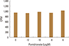
1. In vitro effect of pamidronate in human IVD cells DNA synthesis
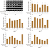
Human IVD cell culture in three dimensional alginate beads with various concentrations of pamidronate (10-12, 10-9, 10-6, and 10-3 M) showed no significant increase in DNA synthesis compared to control culture without pamidronate, indicating there was neither cellular proliferative nor cytotoxic effect of pamidronate on human intervertebral disc cells in vitro (Fig. 1).
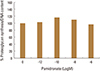
2. Proteoglycan synthesis
Human IVD cell culture in three dimensional alginate beads with low various concentrations of pamidronate (10-12, 10-9, 10-6, and 10-3 M) showed no significant increase in proteoglycan synthesis (Fig. 2).
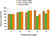
3. Expression of collagen type I, collagen type II, and aggrecan mRNA
In densitometric assay of RT-PCR, human IVD cell culture in three dimensional alginate beads with various dose of pamidronate (10-12, 10-9, 10-6, and 10-3 M) showed no statistically significant changes in mRNA expression of type I collagen, type II collagen, and aggrecan compared to control (Fig. 3).
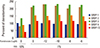
4. The expression of MMP-1, -2, -3, -9, and -13 mRNA in serum starvation
Under the lower serum culture (1% FBS), human IVD cells in alginate beads showed upregulation of MMP-2, -3, -9, and -13 mRNA while no significant change in MMP-1 mRNA expression (Fig. 4).
5 The expression of MMP-1, -2, -3, -9, and -13 mRNA in serum starvation and TNF-α
Human IVD cells in lower serum and TNF-α (10 ng/mL) demonstrated also upregulations of MMP-2, -3, -9, and -13 mRNA. Human IVD cells with various doses of pamidronate (10-12, 10-9, 10-8, and 10-6 M) and lower serum and TNF-α (10 ng/mL) revealed partial down-regulation of MMPs. In detail, human IVD cells with above mentioned condition, showed down-regulation of MMP-1 mRNA (pamidronate 10-12), MMP-2 (pamidronate 10-9), MMP-3 (pamidronate10-8), MMP-9 (pamidronate 10-12,10-10, and 10-8), and MMP-13 (pamidronate 10-12, 10-10, and 10-8) (Fig. 5).
DISCUSSION
BPs have profound effect on skeletal tissue since they have inherent affinity to hydroxyapatite. Fundamental effect of BPs on skeletal tissue is inhibition of osteoclastic bone resorption by increasing apoptosis and decreasing cellular function of osteoclast, leading to maintenance of trabecular and cortical bone volume.[12] Hence BPs have been utilized in various skeletal conditions due to increased osteoclastic activity including postmenopausal osteoporosis, steroid induced osteoporosis, Paget's disease, tumoral bone disease, and particle induced bone resorption around metal implant.[171820] Recently, non-skeletal effects of BPs have been investigated in various tissues i.e., articular cartilage, chondrocyte.[1114192021] With regard to non-skeletal effect of BPs, it appears to be beneficial in terms of metabolism and maintenance of chondrogenic phenotype which include articular cartilage and IVD. IVDs are known to express chondrogenic phenotype and have similar functions like articular cartilage.
In this experimental study, in vitro effects of pamidronate (second generation nitrogen-containing BPs) were investigated using human IVD cells. The results showed that pamidronate exhibited no significant changes on cellular proliferation, proteoglycan synthesis, and chondrogenic phenotypycal expressions which comprise type II collagen and aggrecan mRNA. Also Pamidronate did not exert any cytotoxic effect on human IVD cells as evidence by sustained DNA and proteoglycan synthesis. Those findings imply pamidronate appears to maintain chondrogenic phenotypes at least, not down-regulate expressions of chondrogenic phenotype.
In lower serum condition (FBS 1%), there was significant upregulation of MMP-2, -3, -9, and -13 mRNA expression in human IVD cells. Contrary to that, human IVD cells under lower serum and TNF-α stimulation exhibited demonstrated down-regulation of MMPs in certain dose of pamidronate. Pamidronate with a dose of 10-12 M showed down-regulation of MMP-1, -9, and -13 mRNA, pamidronate with 10-8 M showed down-regulation of MMP-3, -9, and -13 mRNA, and pamidronate with 10-10 M demonstrated down-regulation of MMP-9, and -13 mRNA. Therefore lower dose of pamidronate (10-12 to 10-8 M) inhibited the expression of MMPs in IVD cell cultures with lower serum and TNF-α. Only exception was MMP-2 mRNA expression which was inhibited by pamidronate with a dose of 10-6 M. Synovial fibroblasts in response to TNF-α and interleukins produces cytokines which cause inflammation, angiogenesis, and cartilage degradation through matrix-degrading enzymes including MMP-13.[3334353637] Human IVD cells with response to TNF-α clearly demonstrated that increase in MMPs expression most importantly MMP-13 and those upregulations of MMPs mRNA expression were significantly abrogated with lower dose of pamidronate (10-12 to 10-8 M) in vitro. TNF-α plays an important role in inflammatory arthritis and IVD degeneration.[192122] TNF-α secreted from inflammatory cells and IVD cells triggers MMPs expression which ends up in vicious cycle of cell apoptosis and matrix degradation. The result of the current study demonstrated pamidronate can maintain chondrogenic phenotype and also down-regulated TNF-α induced expressions of MMP-1, -2, -3, -9, and -13. TNF-α blocker is now widely used in the treatment of inflammatory arthritis, sero-negative spondyloarthropathy, and in certain cases of IVD herniation.[38] Cartilageneous matrix protection of pamidronate by maintaining phenotypical expression and inhibiting expression of MMPs might render another beneficial and therapeutic importance in the treatment of IVD degeneration. The limitation of this study is primarily focused on the changes in gene expression and is insufficient in the expression of the protein i.e., MMPs and collagens. Also, it was not demonstrated how pamidronate inhibits TNF-α directly in this study. In the future, it is needed how pamidronate acts at the TNF-α block and inhibits the expression of MMPs in the protein level.
The results of our study correspond with the growing evidence that despite their apoptotic effects on osteoclasts and macrophages, BPs appear to have beneficial effects on other cells. It has been shown that BPs can enhance the differentiation and bone forming activities of osteoblasts [39] and also it has been demonstrated that BPs prevent steroid induced apoptosis of osteoblasts in vitro and in vivo. [40] Furthermore, recent results have clearly demonstrated that pamidronate has chondroprotective effect via maintaining chondrogenic phenotype expression and down-regulating MMPs expression.
In conclusion, pamidronate, nitrogen-containing second generation BPs, was safe in metabolism of IVD in vitro maintaining chondrogenic phenotype and matrix synthesis, and down-regulated TNF-α induced MMPs expression. Hence BPs can exert beneficial and therapeutic effects on IVD.

 XML Download